


Page 475 - Textbook of Pathology, 6th Edition
P. 475
stroma. Occasional multinucleate tumour giant cells are  459
                                                                 present.
                                                                 iii) Numerous capillary-sized blood vessels are found and
                                                                 the tumour cells may be aggregated around them.
                                                                 iv) A few lymphocytes, plasma cells and macrophages are
                                                                 seen.
                                                                 v) Foci of haemorrhage and deposits of haemosiderin
                                                                 granules are often present.

                                                               SECONDARY TUMOURS
                                                               Metastatic tumours of the heart are more common than the
                                                               primary tumours. About 10% cases with disseminated cancer
                                                               have metastases in the heart. Most of these result from
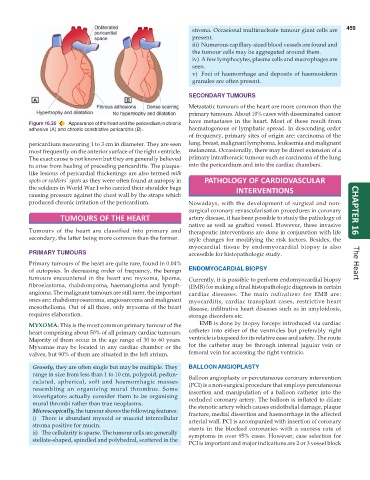
           Figure 16.35  Appearance of the heart and the pericardium in chronic
           adhesive (A) and chronic constrictive pericarditis (B).  haematogenous or lymphatic spread. In descending order
                                                               of frequency, primary sites of origin are: carcinoma of the
           pericardium measuring 1 to 3 cm in diameter. They are seen  lung, breast, malignant lymphoma, leukaemia and malignant
           most frequently on the anterior surface of the right ventricle.  melanoma. Occasionally, there may be direct extension of a
           The exact cause is not known but they are generally believed  primary intrathoracic tumour such as carcinoma of the lung
           to arise from healing of preceding pericarditis. The plaque-  into the pericardium and into the cardiac chambers.
           like lesions of pericardial thickenings are also termed milk
           spots or soldiers’ spots as they were often found at autopsy in  PATHOLOGY OF CARDIOVASCULAR
           the soldiers in World War I who carried their shoulder bags         INTERVENTIONS
           causing pressure against the chest wall by the straps which
           produced chronic irritation of the pericardium.     Nowadays, with the development of surgical and non-
                                                               surgical coronary revascularisation procedures in coronary  CHAPTER 16
                     TUMOURS OF THE HEART                      artery disease, it has been possible to study the pathology of
                                                               native as well as grafted vessel. However, these invasive
           Tumours of the heart are classified into primary and  therapeutic interventions are done in conjunction with life
           secondary, the latter being more common than the former.  style changes for modifying the risk factors. Besides, the
                                                               myocardial tissue by endomyocardial biopsy is also
           PRIMARY TUMOURS                                     accessible for histopathologic study.

           Primary tumours of the heart are quite rare, found in 0.04%                                                The Heart
           of autopsies. In decreasing order of frequency, the benign  ENDOMYOCARDIAL BIOPSY
           tumours encountered in the heart are: myxoma, lipoma,  Currently, it is possible to perform endomyocardial biopsy
           fibroelastoma, rhabdomyoma, haemangioma and lymph-  (EMB) for making a final histopathologic diagnosis in certain
           angioma. The malignant tumours are still rarer, the important  cardiac diseases. The main  indications for EMB are:
           ones are: rhabdomyosarcoma, angiosarcoma and malignant  myocarditis, cardiac transplant cases, restrictive heart
           mesothelioma. Out of all these, only myxoma of the heart  disease, infiltrative heart diseases such as in amyloidosis,
           requires elaboration.                               storage disorders etc.
           MYXOMA. This is the most common primary tumour of the   EMB is done by biopsy forceps introduced via cardiac
           heart comprising about 50% of all primary cardiac tumours.  catheter into either of the ventricles but preferably right
           Majority of them occur in the age range of 30 to 60 years.  ventricle is biopsied for its relative ease and safety. The route
           Myxomas may be located in any cardiac chamber or the  for the catheter may be through internal jugular vein or
           valves, but 90% of them are situated in the left atrium.  femoral vein for accessing the right ventricle.

            Grossly, they are often single but may be multiple. They  BALLOON ANGIOPLASTY
            range in size from less than 1 to 10 cm, polypoid, pedun-  Balloon angioplasty or percutaneous coronary intervention
            culated, spherical, soft and haemorrhagic masses   (PCI) is a non-surgical procedure that employs percutaneous
            resembling an organising mural thrombus. Some      insertion and manipulation of a balloon catheter into the
            investigators actually consider them to be organising  occluded coronary artery. The balloon is inflated to dilate
            mural thrombi rather than true neoplasms.          the stenotic artery which causes endothelial damage, plaque
            Microscopically, the tumour shows the following features:  fracture, medial dissection and haemorrhage in the affected
            i) There is abundant myxoid or mucoid intercellular  arterial wall. PCI is accompanied with insertion of coronary
            stroma positive for mucin.                         stents in the blocked coronaries with a success rate of
            ii) The cellularity is sparse. The tumour cells are generally  symptoms in over 95% cases. However, case selection for
            stellate-shaped, spindled and polyhedral, scattered in the
                                                               PCI is important and major indications are 2 or 3 vessel block

