


Page 95 - Textbook of Pathology, 6th Edition
P. 95
ribonucleoproteins. It is also specific for SLE but is seen in                                           79
           about 25% cases.
           iv) Other non-specific antibodies. Besides above, there are
           several other antibody tests which lack specificity for SLE.
           These are as follows:
           a) Anti-ribonucleoproteins (anti-RNP) seen in 40% cases of SLE                                             CHAPTER 4
           but seen more often in Sjögren’s syndrome.
           b) Anti-histone antibody, which is antibody against histone
           associated with DNA in chromatin, is seen particularly in
           cases of drug-induced lupus than in SLE.
           c) Antiphospholipid antibodies or lupus anticoagulant are tests
           for thrombotic complications in cases of SLE.
           d) Antiribosomal P antibody  is antibody against protein in
           ribosomes and is seen in CNS lupus.
              The source of these autoantibodies as well as hyper-
           gammaglobulinaemia seen in SLE is the polyclonal activa-
           tion of B cells brought about by following derangements:
           1. Immunologic factors. These include:
           i) an inherited defect in B cells;
           ii) stimulation of B cells by micro-organisms;
           iii) T helper cell hyperactivity; and                                                                      Immunopathology Including Amyloidosis
           iv) T suppressor cell defect.
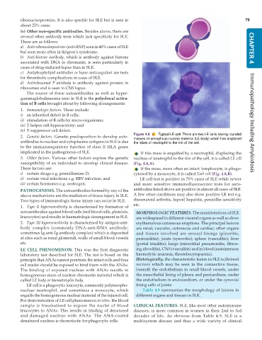
                                                               Figure 4.8  Typical LE cell. There are two LE cells having rounded
           2. Genetic factors. Genetic predisposition to develop auto-  masses of amorphous nuclear material (LE body) which has displaced
           antibodies to nuclear and cytoplasmic antigens in SLE is due  the lobes of neutrophil to the rim of the cell.
           to the immunoregulatory function of class II HLA genes
           implicated in the pathogenesis of SLE.                  If this mass is engulfed by a neutrophil, displacing the
           3. Other factors. Various other factors express the genetic  nucleus of neutrophil to the rim of the cell, it is called LE cell
           susceptibility of an individual to develop clinical disease.  (Fig. 4.8,A).
           These factors are:                                      If the mass, more often an intact lymphocyte, is phago-
           i) certain drugs e.g. penicillamine D;              cytosed by a monocyte, it is called Tart cell (Fig. 4.8,B).
           ii) certain viral infections e.g. EBV infection; and   LE cell test is positive in 70% cases of SLE while newer
           iii) certain hormones e.g. oestrogen.               and more sensitive immunofluorescence tests for auto-
           PATHOGENESIS. The autoantibodies formed by any of the  antibodies listed above are positive in almost all cases of SLE.
           above mechanisms are the mediators of tissue injury in SLE.  A few other conditions may also show positive LE test e.g.
           Two types of immunologic tissue injury can occur in SLE:  rheumatoid arthritis, lupoid hepatitis, penicillin sensitivity
           1. Type II hypersensitivity is characterised by formation of  etc.
           autoantibodies against blood cells (red blood cells, platelets,  MORPHOLOGIC FEATURES. The manifestations of SLE
           leucocytes) and results in haematologic derangement in SLE.  are widespread in different visceral organs as well as show
           2. Type III hypersensitivity is characterised by antigen-anti-  erythematous cutaneous eruptions. The principal lesions
           body complex (commonly DNA-anti-DNA antibody;         are renal, vascular, cutaneous and cardiac; other organs
           sometimes Ig-anti-Ig antibody complex) which is deposited  and tissues involved are serosal linings (pleuritis,
           at sites such as renal glomeruli, walls of small blood vessels  pericarditis); joints (synovitis); spleen (vasculitis); liver
           etc.                                                  (portal triaditis); lungs (interstitial pneumonitis, fibros-
           LE CELL PHENOMENON. This was the first diagnostic     ing alveolitis), CNS (vasculitis) and in blood (autoimmune
           laboratory test described for SLE. The test is based on the  haemolytic anaemia, thrombocytopaenia).
           principle that ANAs cannot penetrate the intact cells and thus  Histologically, the characteristic lesion in SLE is fibrinoid
           cell nuclei should be exposed to bind them with the ANAs.  necrosis which may be seen in the connective tissue,
           The binding of exposed nucleus with ANAs results in   beneath the endothelium in small blood vessels, under
           homogeneous mass of nuclear chromatin material which is  the mesothelial lining of pleura and pericardium, under
           called LE body or haematoxylin body.                  the endothelium in endocardium, or under the synovial
              LE cell is a phagocytic leucocyte, commonly polymorpho-  lining cells of joints.
           nuclear neutrophil, and sometimes a monocyte, which      Table 4.9 summarises the morphology of lesions in
           engulfs the homogeneous nuclear material of the injured cell.  different organs and tissues in SLE.
           For demonstration of LE cell phenomenon in vitro, the blood
           sample is traumatised to expose the nuclei of blood  CLINICAL FEATURES. SLE, like most other autoimmune
           leucocytes to ANAs. This results in binding of denatured  diseases, is more common in women in their 2nd to 3rd
           and damaged nucleus with ANAs. The ANA-coated       decades of life. As obvious from Table 4.9, SLE is a
           denatured nucleus is chemotactic for phagocytic cells.  multisystem disease and thus a wide variety of clinical

