


Page 100 - Textbook of Pathology, 6th Edition
P. 100
84


     SECTION I


     General Pathology and Basic Techniques


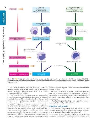
           Figure 4.10  Pathogenesis of two main forms of amyloid deposition (AL = Amyloid light chain; AA = Amyloid-associated protein; GAG =
           glycosaminoglycan; AP = Amyloid P component). The sequence on left shows general schematic representation common to both major forms of
           amyloidogenesis.


           1. Pool of amyloidogenic precursor  protein is present in  haemodialysis) and prionosis (in which β-pleated sheet is
           circulation in different clinical settings and in response to  formed de novo).
           stimuli e.g. increased hepatic synthesis of AA or ATTR,  5. The role of non-fibrillar components such as AP, apoE and
           increased synthesis of AL etc.                      GAGs in amyloidosis is unclear; probably they facilitate in
           2. A nidus for fibrillogenesis, meaning thereby an alteration  aggregation of proteins and protein folding leading to fibril
           in microenvironment, to stimulate deposition of amyloid  formation, substrate adhesion and protection from
           protein is formed. This alteration involves changes and  degradation.
           interaction between basement membrane proteins  and    Based on this general pathogenesis, deposition of AL and
           amyloidogenic protein.                              AA amyloid is briefly outlined below:
           3. Partial degradation or proteolysis occurs prior to deposition
           of fibrillar protein which may occur in macrophages or  Deposition of AL Amyloid
           reticuloendothelial cells e.g. partial degradation of AL, AA.  1. The stimulus for production of AL amyloid is some
           4. Exceptions to this generalisation, however, are seen in ATTR  disorder of immunoglobulin synthesis e.g. multiple myeloma,
           (heredofamilial type in which there are amino acid mutations  B cell lymphoma, other plasma cell dyscrasias.
           in most cases), Aβ2M (in which there are elevated levels of  2. Excessive immunoglobulin production is in the form of
           normal  β2M protein which remain unfiltered during  monoclonal gammopathy i.e. there is production of either intact

