


Page 90 - Textbook of Pathology, 6th Edition
P. 90
74
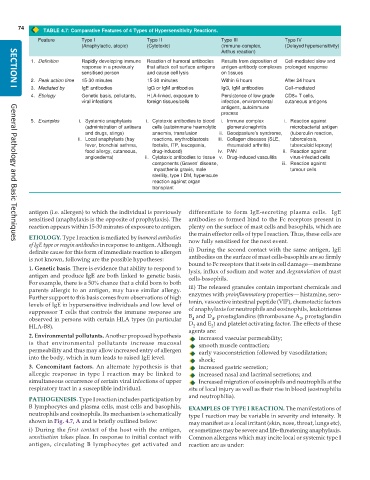
TABLE 4.7: Comparative Features of 4 Types of Hypersensitivity Reactions.
Feature Type I Type II Type III Type IV
(Anaphylactic, atopic) (Cytotoxic) (Immune-complex, (Delayed hypersensitivity)
Arthus reaction)
1. Definition Rapidly developing immune Reaction of humoral antibodies Results from deposition of Cell-mediated slow and
response in a previously that attack cell surface antigens antigen-antibody complexes prolonged response
sensitised person and cause cell lysis on tissues
2. Peak action time 15-30 minutes 15-30 minutes Within 6 hours After 24 hours
3. Mediated by IgE antibodies IgG or IgM antibodies IgG, IgM antibodies Cell-mediated
SECTION I
4. Etiology Genetic basis, pollutants, HLA-linked, exposure to Persistence of low grade CD8+ T cells,
viral infections foreign tissues/cells infection, environmental cutaneous antigens
antigens, autoimmune
process
5. Examples i. Systemic anaphylaxis i. Cytotoxic antibodies to blood i. Immune complex i. Reaction against
(administration of antisera cells (autoimmune haemolytic glomerulonephritis microbacterial antigen
and drugs, stings) anaemia, transfusion ii. Goodpasture’s syndrome, (tuberculin reaction,
ii. Local anaphylaxis (hay reactions, erythroblastosis iii. Collagen diseases (SLE, tuberculosis,
fever, bronchial asthma, foetalis, ITP, leucopenia, rheumatoid arthritis) tuberculoid leprosy)
food allergy, cutaneous, drug-induced) iv. PAN ii. Reaction against
angioedema) ii. Cytotoxic antibodies to tissue v. Drug-induced vasculitis virus-infected cells
components (Graves’ disease, iii. Reaction against
myasthenia gravis, male tumour cells
sterility, type I DM, hyperacute
reaction against organ
transplant
antigen (i.e. allergen) to which the individual is previously differentiate to form IgE-secreting plasma cells. IgE
sensitised (anaphylaxis is the opposite of prophylaxis). The antibodies so formed bind to the Fc receptors present in
reaction appears within 15-30 minutes of exposure to antigen. plenty on the surface of mast cells and basophils, which are
the main effector cells of type I reaction. Thus, these cells are
ETIOLOGY. Type I reaction is mediated by humoral antibodies now fully sensitised for the next event.
General Pathology and Basic Techniques
of IgE type or reagin antibodies in response to antigen. Although
definite cause for this form of immediate reaction to allergen ii) During the second contact with the same antigen, IgE
is not known, following are the possible hypotheses: antibodies on the surface of mast cells-basophils are so firmly
bound to Fc receptors that it sets in cell damage—membrane
1. Genetic basis. There is evidence that ability to respond to lysis, influx of sodium and water and degranulation of mast
antigen and produce IgE are both linked to genetic basis. cells-basophils.
For example, there is a 50% chance that a child born to both
parents allergic to an antigen, may have similar allergy. iii) The released granules contain important chemicals and
Further support to this basis comes from observations of high enzymes with proinflammatory properties— histamine, sero-
levels of IgE in hypersensitive individuals and low level of tonin, vasoactive intestinal peptide (VIP), chemotactic factors
suppressor T cells that controls the immune response are of anaphylaxis for neutrophils and eosinophils, leukotrienes
4
4
observed in persons with certain HLA types (in particular B and D , prostaglandins (thromboxane A , prostaglandin
2
2
2
HLA-B8). D and E ) and platelet activating factor. The effects of these
agents are:
2. Environmental pollutants. Another proposed hypothesis increased vascular permeability;
is that environmental pollutants increase mucosal smooth muscle contraction;
permeability and thus may allow increased entry of allergen early vasoconstriction followed by vasodilatation;
into the body, which in turn leads to raised IgE level. shock;
3. Concomitant factors. An alternate hypothesis is that increased gastric secretion;
allergic response in type I reaction may be linked to increased nasal and lacrimal secretions; and
simultaneous occurrence of certain viral infections of upper Increased migration of eosinophils and neutrophils at the
respiratory tract in a susceptible individual. site of local injury as well as their rise in blood (eosinophilia
and neutrophilia).
PATHOGENESIS. Type I reaction includes participation by
B lymphocytes and plasma cells, mast cells and basophils, EXAMPLES OF TYPE I REACTION. The manifestations of
neutrophils and eosinophils. Its mechanism is schematically type I reaction may be variable in severity and intensity. It
shown in Fig. 4.7, A and is briefly outlined below: may manifest as a local irritant (skin, nose, throat, lungs etc),
i) During the first contact of the host with the antigen, or sometimes may be severe and life-threatening anaphylaxis.
sensitisation takes place. In response to initial contact with Common allergens which may incite local or systemic type I
antigen, circulating B lymphocytes get activated and reaction are as under:

