


Page 98 - Textbook of Pathology, 6th Edition
P. 98
82                                                      II. Non-fibrillar components  which include  P-component
                            AMYLOIDOSIS
                                                               predominantly; there are several different proteins which
           Amyloidosis is the term used for a group of diseases charac-  together constitute the remaining 5% of amyloid.
           terised by extracellular deposition of fibrillar proteinaceous
           substance called amyloid having common morphological  I. Fibril Proteins
           appearance, staining properties and physical structure but
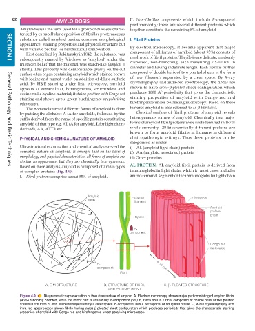
           with variable protein (or biochemical) composition.  By electron microscopy, it became apparent that major
              First described by Rokitansky in 1842, the substance was  component of all forms of amyloid (about 95%) consists of
           subsequently named by Virchow as ‘amyloid’ under the  meshwork of fibril proteins. The fibrils are delicate, randomly
     SECTION I
           mistaken belief that the material was starch-like (amylon =  dispersed, non-branching, each measuring 7.5-10 nm in
           starch). This property was demonstrable grossly on the cut  diameter and having indefinite length. Each fibril is further
           surface of an organ containing amyloid which stained brown  composed of double helix of two pleated sheets in the form
           with iodine and turned violet on addition of dilute sulfuric  of  twin filaments separated by a clear space. By X-ray
           acid. By  H&E staining under  light microscopy, amyloid  crystallography and infra-red spectroscopy, the fibrils are
           appears as extracellular, homogeneous, structureless and  shown to have  cross-β-pleated  sheet configuration which
           eosinophilic hyaline material; it stains positive with Congo red  produces 1000 A° periodicity that gives the characteristic
                                                               staining properties of amyloid with Congo red and
           staining and shows apple-green birefringence on polarising  birefringence under polarising microscopy. Based on these
           microscopy.
              The nomenclature of different forms of amyloid is done  features amyloid is also referred to as β-fibrillosis.
           by putting the alphabet A (A for amyloid), followed by the  Chemical analysis of fibril proteins of amyloid reveals
           suffix derived from the name of specific protein constituting  heterogeneous nature of amyloid. Chemically two major
           amyloid of that type e.g. AL (A for amyloid, L for light chain-  forms of amyloid fibril proteins were first identified in 1970s
           derived), AA, ATTR etc.                             while currently  20 biochemically different proteins are
                                                               known to form amyloid fibrils in humans in different
           PHYSICAL AND CHEMICAL NATURE OF AMYLOID             clinicopathologic settings. Thus these proteins can be
                                                               categorised as under:
           Ultrastructural examination and chemical analysis reveal the  i) AL (amyloid light chain) protein
           complex nature of amyloid.  It emerges that on the basis of  ii) AA (amyloid associated) protein
           morphology and physical characteristics, all forms of amyloid are  iii) Other proteins
           similar in appearance, but they are chemically heterogeneous.
           Based on these analysis, amyloid is composed of 2 main types  AL PROTEIN.  AL amyloid fibril protein is derived from
     General Pathology and Basic Techniques
           of complex proteins (Fig. 4.9):                     immunoglobulin light chain, which in most cases includes
           I. Fibril proteins comprise about 95% of amyloid.   amino-terminal segment of the immunoglobulin light chain


           Figure 4.9  Diagrammatic representation of the ultrastructure of amyloid. A, Electron microscopy shows major part consisting of amyloid fibrils
           (95%) randomly oriented, while the minor part is essentially P-component (5%) B, Each fibril is further composed of double helix of two pleated
           sheets in the form of twin filaments separated by a clear space. P-component has a pentagonal or doughnut profile. C, X-ray crystallography and
           infra-red spectroscopy shows fibrils having cross-β-pleated sheet configuration which produces periodicity that gives the characteristic staining
           properties of amyloid with Congo red and birefringence under polarising microscopy.

