Page 56 - PRE-U STPM BIOLOGY TERM 1
P. 56

Biology Term 1  STPM  Chapter 2 Structure of Cells and Organelles

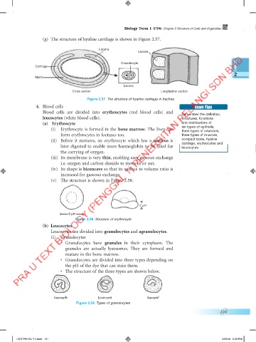
                     (g)  The structure of hyaline cartilage is shown in Figure 2.57.

                                               Lacuna
                                                                 Lacuna

                                                         Chondrocyte
                 Cartilage
                                                                                                              2
                 Matrix
                                                           Lacuna
                                   Cross section                            Longitudinal section
                                          Figure 2.57  The structure of hyaline cartilage in trachea
                  4.  Blood cells                                                           Exam Tips
                     Blood  cells are divided into  erythrocytes  (red blood cells) and   Remember the definition,
                     leucocytes (white blood cells).                                  structures, functions
                     (a)   Erythrocyte                                                and distributions of
                         (i)  Erythrocyte is formed in the bone marrow. The liver can   six types of epithelia,
                                                                                      three types of neurones,
                             form erythrocytes in foetuses too.                       three types of muscles,
                         (ii)  Before it matures, an erythrocyte which has a  nucleus  is   compact bone, hyaline
                             later digested to enable more haemoglobin to be filled for   cartilage, erythrocytes and
                                                                                      leucocytes.
                             the carrying of oxygen.
                         (iii)  Its membrane is very thin, enabling easy gaseous exchange
                             i.e. oxygen and carbon dioxide to move in or out.
                         (iv)  Its shape is biconcave so that its surface to volume ratio is
                             increased for gaseous exchange.
                         (v)  The structure is shown in Figure 2.58.


                                                                  2 μm

                                 8 μm
                                    Figure 2.58  Structure of erythrocyte
                     (b)  Leucocytes
                         Leucocytes are divided into granulocytes and agranulocytes.
                         (i)  Granulocytes
                             •  Granulocytes  have  granules  in  their cytoplasm.  The
                               granules are actually lysosomes. They are formed and
                               mature in the bone marrow.
                             •  Granulocytes are divided into three types depending on
                               the pH of the dye that can stain them.
                             •  The structure of the three types are shown below.


                          Neutrophill          Eosinophil            Basophill
                                                                      Basophil
                          Neutrophil
                                     Figure 2.59  Types of granulocytes
                                                                                                        101


         02[STPM Bio T1].indd   101                                                                     3/29/18   5:08 PM
   51   52   53   54   55   56   57   58   59   60   61