
Page 28 - TI Journal 18-1
P. 28
22 BAKER ET AL.
curvature of white matter pathways using in vivo DTI Scalar Metrics
imaging (17). This method is highly sensitive to white A symmetric 3x3 diffusion tensor characterizes
matter changes within entire tracts and, therefore, water diffusion in brain tissues. This model represents
may be more advantageous than methods that involve the diffusion pattern with a second-order tensor that
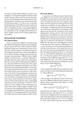
placing regions of interest on two-dimensional scalar can be decomposed into three non-negative eigen-
DTI parameter maps (17). In this review, we describe values and three eigenvectors that describe the mag-
the fundamentals of the diffusion tensor model and nitude and orientation of water diffusion in each
qtDTI technology. We then review the existing liter- voxel (Figure 1). Eigenvalues describe the shape and
ature on length-based metrics using qtDTI, followed size of the tensor, independent of orientation, while
by a discussion of the strengths and limitations of eigenvectors describe the orientation of the tensor,
qtDTI. Finally, a brief review of future applications independent of shape and size. The tensor model
is provided. parameterizes the diffusion in each voxel with an
ellipsoid whose diameter in any direction estimates
DIFFUSION MR TECHNIQUES the diffusivity in that direction and whose major prin-
DTI Physical Basis ciple axis is oriented in the direction of maximum
DTI is a noninvasive magnetic resonance imag- diffusivity. The major axis of the ellipsoid (v1) points
ing (MRI) technology that measures water diffusion in the direction of the maximum diffusivity (λ1) of
at each voxel in the brain. Water molecules diffuse a voxel. The direction of the maximum diffusion is
differently along tissues depending on tissue micro- oriented in the direction of the major fiber tract in
structure and the presence of anatomical barriers. the voxel. The directions perpendicular to the main
One simple and useful way to characterize diffusion fiber orientation along the medium (v 2) and minor
at a location in the brain is along a spectrum between axes (v3) of the diffusion ellipsoid are also computed
isotropic and anisotropic. Diffusion that is highly (λ2, λ3) in the tensor analysis. DTI scalar metrics are
similar in all directions (i.e., isotropic diffusion) is functions of three diffusion eigenvalues (λ1, λ2, λ3).
typically observed in grey matter and cerebrospinal Axial diffusivity (AD = λ1) is the maximum diffu-
fluid. By contrast, directionally dependent diffusion sivity in the voxel and decreases with greater axonal
(i.e., anisotropic diffusion) is observed in white matter injury (15,29). Radial diffusivity
due to the linear organization of the fiber tracts. Water
within these tracts preferentially diffuses in one direc-
tion because physical barriers such as axonal walls is the average of the diffusivity perpendicular to the
and myelin restrict water movement in other direc- major axis and increases with abnormal myelination
tions (5,24,47,48). Neuropathological mechanisms (1). Mean diffusivity
associated with multiple conditions, including sub-
cortical ischemia, neurodegeneration, and traumatic
brain injury, cause reductions in the linear organi- is the average of the diffusivity values of the three
zation of white matter pathways with corresponding axes of the diffusion ellipsoid and is sensitive to cel-
reductions in linear anisotropy (5,19,48,52). DTI is lularity, edema, and necrosis (46) (Figure 2). Lastly,
sensitive to these changes in linear anisotropy even fractional anisotropy
when white matter integrity appears healthy based
on structural neuroimaging methods (referenced as
normal appearing white matter) (4,30), making DTI parameterizes the degree to which the diffusion
a powerful in vivo imaging method for the examina- ellipsoid deviates from spherical. FA is a normal-
tion of the microstructural integrity of white matter. ized measure ranging from zero to one that decreases
with axonal degeneration, abnormal myelination, and
fiber orientation dispersion (27, 35-36. 47) (Figure
2). These scalar metrics describe microstructural