
Page 36 - Human Environment Interface (4)
P. 36
Multi-Interface Domain Analysis
Figure 1. Multi-interface domain illustration. Domain a of protein proteins are either at the protein level (e.g., protein-protein
A interacting with domain b of protein B produces interface a–b on interaction, protein complex identification) [7,8], or at the domain
domain a, and domain a binding to protein C generates interface a–c level (e.g., domain-domain interaction, domain transitivity analysis)
on domain a. Interfaces a–b and a–c are distinguishable on domain a, [9,10], or at residue level (e.g., interface residue identification, hot
thus domain a is a multi-interface domain. spots prediction) [11,12]. Studies are seldom undertaken at the
doi:10.1371/journal.pone.0050821.g001 interface level, which is in the middle between the domain and
residue levels. This is probably attributable to uncertainties in
The multiple interfaces in a domain can be grouped into subsets locating an interface due to the adaptation, context-awareness, or
such that each of them share a unique biological function. These re-configuration of interfaces [6,13,14], in contrast to the clear
functions are usually distinguishable and non exchangeable. As an boundaries possessed by a protein, domain or residue. Some
example shown in Figure 2, the three functions of the plasmin notable exceptions are the works of identifying conserved interface
catalytic domain cannot be swapped with regard to their patterns by interface alignment [15], detecting common 3D sites in
interfaces. More interestingly, sometimes the number of such proteins by frequent graph patterns of stereochemical atom groups
subsets of the interfaces in a domain can be large. As protein [16], uncovering functional sites in protein families by recurring
functions are currently annotated at the domain level, such as graph patterns [17], and delineating biological functions of
those by GO [5], it is difficult to figure out which interface in a proteins by common atomic motifs of interfaces [18,19]. However,
domain possesses what function based on the current annotations. all these methods mix up interfaces from different domains even if
The study of multi-interface domains can associate interfaces with they are remarkably different. In addition, multiple interfaces in
their functions more precisely. Yet almost all past studies of one domain are deemed as independent as no relation has been
unveiled. Further more, associations between multiple interfaces
and their biological functions still remain unanswered.
In this study, we address the following questions: (i) What kind
of domains prefer the multi-interface property? That is, we want to
know the distribution of domains that have multiple interfaces. (ii)
What are the fingerprints of an interface, or a subset of interfaces,
in a domain? That is, we want to discover unique structures in a
domain that distinguish the multiple interfaces from each other.
(iii) What are the relationships between the multiple interfaces in a
domain? That is, we want to see whether the multiple interfaces in
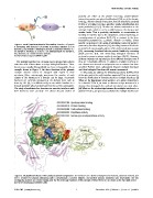
Figure 2. Multiple interfaces in the catalytic domain of plasmin. The interfaces are colored in limegreen (46 residues), marine (6 residues), and
red (7 residues) for plasmin interacting with a streptokinase, a protein inhibitor, and another plasmin symmetric unit, respectively. The two
overlapping residues are colored orange, and the five molecular functions of this domain retrieved from GO are shown at the top right corner.
doi:10.1371/journal.pone.0050821.g002
PLOS ONE | www.plosone.org 2 December 2012 | Volume 7 | Issue 12 | e50821