
Page 41 - Human Environment Interface (4)
P. 41
Multi-Interface Domain Analysis
Table 4. Top ten multi-interface domains with the largest numbers of proteins.
Domain name # multi-interface proteins avg # interfaces
Ig heavy chain variable domain, VH 43 2.4(+0.7)
Ig light chain k variable domain, VL-k 35 2.5(+0.8)
Ig heavy chain c constant domain 1, CH1-c 28 2.3(+0.6)
Ig light chain k constant domain, CL-k 18 2.4(+0.6)
Hemoglobin, beta-chain 17 2.8(+0.9)
Proteasome beta subunit (catalytic) 17 7.6(+3.2)
T-cell antigen receptor 16 3.1(+1.6)
Hemoglobin, alpha-chain 14 3.1(+0.5)
Nucleoside diphosphate kinase, NDK 13 2.9(+0.3)
Dodecameric ferritin homolog 12 5.4(+1.4)
doi:10.1371/journal.pone.0050821.t004
interface proteins, we cannot get reasonable results from these two besides a sufficient number of multi-interface proteins holding this
types of interactions. domain.
Analysis on multiple interfaces within the same domain Fingerprints of interface. Given a set of multiple interfaces
The top ten multi-interface domains with the largest numbers of in a domain, we fish out interface-specific fingerprints by the
mining of closed frequent subgraphs (substructures) from the
proteins are shown in Table 4. Unexpectedly, domains from the corresponding interface graph database. These frequent substruc-
immune system have the most number of multi-interface proteins. tures capture the natural organizations of interface residues. In the
This observation in part can be explained by the fact that a small past, frequent sub-structures have been successfully applied to
portion of hypervariable regions exist in these proteins [48]. That study protein structure and function [49].
is, although these proteins have the same domain, they do contain
different interfaces due to mutations occurring in the hypervari- Our experiments on identifying interface fingerprints are
able regions. carried out on Ig VH domain and proteasome beta subunit
domain separately. Generally, we obtained a great number of non-
Table 4 also unveils the wide coverage of biological functions trivial closed frequent substructures for each interface of the two
played by some domain. For example, the proteasome beta domains. Full results are provided in Supplement Data S1. We
subunit (catalytic) domain has around seven different interfaces then examine whether these graph patterns (fingerprints) are
which are involved in different biological processes. This is quite domain-specific or not. To this end, we compared fingerprints
different from the Ig VH domain with the typical two interfaces between interfaces in various dimensions. First, we directly
playing the Ig VL protein binding role and the role of antigen compared residue composition of different interfaces. The detailed
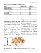
recognition. results are shown in Figure 5 and Figure 6 for Ig VH domain and
proteasome beta subunit domain, respectively. It is obvious that
Since there are as many as 1,730 multi-interface domains—see residue preferences are divergent for different interfaces. Second,
Figure 4 and Table 2—and exploring properties of every multi- we explored fingerprint isomorphism between different interfaces.
interface domain is not our purpose in this study, we undertake Not surprisingly, we identified just one isomorphic fingerprint
analysis on two domains: Ig VH domain and proteasome beta between the interfaces of Ig VH domain and very few isomorphic
subunit domain. The former is contained in the largest number of fingerprints between interfaces of proteasome beta subunit domain
multi-interface proteins and the latter has many binding sites
Figure 5. Amino acid distribution of the antigen-binding interface and the protein-binding interface in the Ig VH domain.
doi:10.1371/journal.pone.0050821.g005
PLOS ONE | www.plosone.org 7 December 2012 | Volume 7 | Issue 12 | e50821