
Page 169 - First Aid for the USMLE Step 1 2020, Thirtieth edition [MedicalBooksVN.com]_Neat
P. 169
Microbiology ` microbiology—basic bacteriology Microbiology ` microbiology—basic bacteriology SEcTioN ii 125
Stains
Gram stain First-line lab test in bacterial identification. Bacteria with thick peptidoglycan layer retain crystal
violet dye (gram ⊕); bacteria with thin peptidoglycan layer turn red or pink (gram ⊝) with
counterstain.
These bugs do not Gram stain well (These Little Microbes May Unfortunately Lack Real Color
But Are Everywhere):
Treponema, Leptospira Too thin to be visualized
Mycobacteria Cell wall has high lipid content
Mycoplasma, Ureaplasma No cell wall
Legionella, Rickettsia, Chlamydia, Bartonella, Primarily intracellular; also, Chlamydia lack
Anaplasma, Ehrlichia classic peptidoglycan because of muramic
acid
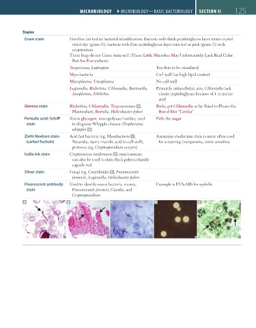
Giemsa stain Rickettsia, Chlamydia, Trypanosomes A , Ricky got Chlamydia as he Tried to Please the
Plasmodium, Borrelia, Helicobacter pylori Bored Hot “Geisha”
Periodic acid–Schiff Stains glycogen, mucopolysaccharides; used PaSs the sugar
stain to diagnose Whipple disease (Tropheryma
whipplei B )
Ziehl-Neelsen stain Acid-fast bacteria (eg, Mycobacteria C , Auramine-rhodamine stain is more often used
(carbol fuchsin) Nocardia; stains mycolic acid in cell wall); for screening (inexpensive, more sensitive)
protozoa (eg, Cryptosporidium oocysts)
India ink stain Cryptococcus neoformans D; mucicarmine
can also be used to stain thick polysaccharide
capsule red
Silver stain Fungi (eg, Coccidioides E , Pneumocystis
jirovecii), Legionella, Helicobacter pylori
Fluorescent antibody Used to identify many bacteria, viruses, Example is FTA-ABS for syphilis
stain Pneumocystis jirovecii, Giardia, and
Cryptosporidium
A B C D E
FAS1_2019_03-Microbiology.indd 125 11/14/19 12:19 PM