
Page 402 - First Aid for the USMLE Step 1 2020, Thirtieth edition [MedicalBooksVN.com]_Neat
P. 402
358 seCtion iii Gastrointestinal ` gastrointestinal—embryology Gastrointestinal ` gastrointestinal—embryology
` gastrointestinal—embryology
Normal Foregut—esophagus to duodenum at level of pancreatic duct and common bile duct insertion
gastrointestinal (ampulla of Vater).
embryology Midgut—lower duodenum to proximal 2/3 of transverse colon.
Hindgut—distal 1/3 of transverse colon to anal canal above pectinate line.
Midgut development:
6th week—physiologic herniation of midgut through umbilical ring
10th week—returns to abdominal cavity + rotates around superior mesenteric artery (SMA),
total 270° counterclockwise
Ventral wall defects Developmental defects due to failure of rostral fold closure (eg, sternal defects [ectopia cordis]),
lateral fold closure (eg, omphalocele, gastroschisis), or caudal fold closure (eg, bladder exstrophy).
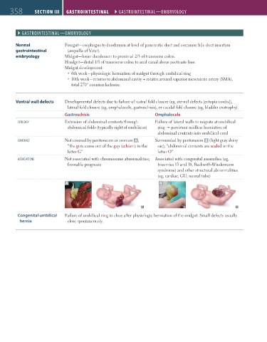
Gastroschisis Omphalocele
etiology Extrusion of abdominal contents through Failure of lateral walls to migrate at umbilical
abdominal folds (typically right of umbilicus) ring persistent midline herniation of
abdominal contents into umbilical cord
CoVerage Not covered by peritoneum or amnion A ; Surrounded by peritoneum B (light gray shiny
“the guts come out of the gap (schism) in the sac); “abdominal contents are sealed in the
letter G” letter O”
assoCiations Not associated with chromosome abnormalities; Associated with congenital anomalies (eg,
favorable prognosis trisomies 13 and 18, Beckwith-Wiedemann
syndrome) and other structural abnormalities
(eg, cardiac, GU, neural tube)
A B
Congenital umbilical Failure of umbilical ring to close after physiologic herniation of the midgut. Small defects usually
hernia close spontaneously.
FAS1_2019_09-Gastrointestinal.indd 358 11/7/19 4:42 PM