
Page 403 - First Aid for the USMLE Step 1 2020, Thirtieth edition [MedicalBooksVN.com]_Neat
P. 403
Gastrointestinal ` gastrointestinal—embryology Gastrointestinal ` gastrointestinal—embryology seCtion iii 359
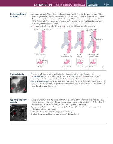
Tracheoesophageal Esophageal atresia (EA) with distal tracheoesophageal fistula (TEF) is the most common (85%)
anomalies and often presents as polyhydramnios in utero (due to inability of fetus to swallow amniotic fluid).
Neonates drool, choke, and vomit with first feeding. TEFs allow air to enter stomach (visible on
CXR). Cyanosis is 2° to laryngospasm (to avoid reflux-related aspiration). Clinical test: failure to
pass nasogastric tube into stomach.
In H-type, the fistula resembles the letter H. In pure EA, CXR shows gasless abdomen.
Trachea Esophagus Tracheoesophageal
fistula
Esophageal
atresia
Normal anatomy Pure EA Pure TEF EA with distal TEF
(atresia or stenosis) (H-type) (most common)
Gastric
bubble
Normal Gasless stomach Prominent gastric bubble
Intestinal atresia Presents with bilious vomiting and abdominal distension within first 1–2 days of life.
Duodenal atresia—failure to recanalize. Abdominal x-ray A shows “double bubble” (dilated
A
stomach, proximal duodenum). Associated with Down syndrome.
Jejunal and ileal atresia—disruption of mesenteric vessels (typically SMA) ischemic necrosis of
fetal intestine segmental resorption: bowel becomes discontinuous. X-ray shows dilated loops of
small bowel with air-fluid levels.
Hypertrophic pyloric Most common cause of gastric outlet obstruction in infants (1:600). Palpable olive-shaped mass in
stenosis epigastric region, visible peristaltic waves, and nonbilious projectile vomiting at ∼ 2–6 weeks old.
More common in firstborn males; associated with exposure to macrolides.
A
Results in hypokalemic hypochloremic metabolic alkalosis (2° to vomiting of gastric acid and
stomach
pyloric wall subsequent volume contraction).
Ultrasound shows thickened and lengthened pylorus A .
pyloric channel Treatment: surgical incision of pyloric muscles (pyloromyotomy).
FAS1_2019_09-Gastrointestinal.indd 359 11/7/19 4:42 PM