Page 411 - First Aid for the USMLE Step 1 2020, Thirtieth edition [MedicalBooksVN.com]_Neat
P. 411

Gastrointestinal  ` gastrointestinal—anatomy   Gastrointestinal  ` gastrointestinal—anatomy           seCtion iii      367


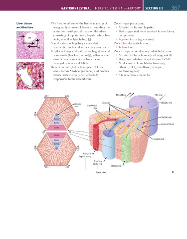
                  Liver tissue           The functional unit of the liver is made up of   Zone I—periportal zone:
                  architecture            hexagonally arranged lobules surrounding the     ƒ Affected 1st by viral hepatitis
                                          central vein with portal triads on the edges     ƒ Best oxygenated, most resistant to circulatory
                   A
                                          (consisting of a portal vein, hepatic artery, bile   compromise
                                          ducts, as well as lymphatics)  A .          ƒ Ingested toxins (eg, cocaine)
                                         Apical surface of hepatocytes faces bile   Zone II—intermediate zone:
                                          canaliculi. Basolateral surface faces sinusoids.     ƒ Yellow fever
                                         Kupffer cells (specialized macrophages) located   Zone III—pericentral vein (centrilobular) zone:
                                          in sinusoids (black arrows in  B ; yellow arrows     ƒ Affected 1st by ischemia (least oxygenated)
                                          show hepatic venule) clear bacteria and     ƒ High concentration of cytochrome P-450
                   B                      damaged or senescent RBCs.                  ƒ Most sensitive to metabolic toxins (eg,
                                         Hepatic stellate (Ito) cells in space of Disse   ethanol, CCl , halothane, rifampin,
                                                                                                4
                                          store vitamin A (when quiescent) and produce   acetaminophen)
                                          extracellular matrix (when activated).      ƒ Site of alcoholic hepatitis
                                          Responsible for hepatic fibrosis.


                                                                                    Blood flow                Bile flow


                                                                          Sinusoids                              Hepatic vein
                                                        Hepatic
                                                        vein    Endothelial
                                                                    cells

                                                                                                                 Stellate cell
                                           Zone 3
                                           Zone 2
                                                                                                                 Space of Disse
                                           Zone 1

                                           Zone 1
                                                                                                             Kup er cell
                                           Zone 2
                                           Zone 3


                                                              Branch of
                                                           hepatic artery
                                                                     Branch of
                                                                    portal vein
                                                                                Bile ductule
                                                                      Portal triad


          FAS1_2019_09-Gastrointestinal.indd   367                                                                      11/7/19   4:42 PM
   406   407   408   409   410   411   412   413   414   415   416