Page 412 - First Aid for the USMLE Step 1 2020, Thirtieth edition [MedicalBooksVN.com]_Neat
P. 412

368        seCtion iii    Gastrointestinal  ` gastrointestinal—anatomy                                                                                        Gastrointestinal  ` gastrointestinal—anatomy


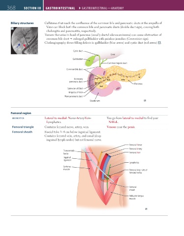
               Biliary structures    Gallstones that reach the confluence of the common bile and pancreatic ducts at the ampulla of
                                       Vater can block both the common bile and pancreatic ducts (double duct sign), causing both
               A
                                       cholangitis and pancreatitis, respectively.
                                     Tumors that arise in head of pancreas (usually ductal adenocarcinoma) can cause obstruction of
                           Endoscope  Cholangiography shows filling defects in gallbladder (blue arrow) and cystic duct (red arrow)  A .
                         CHD           common bile duct Ž enlarged gallbladder with painless jaundice (Courvoisier sign).
                             Pancreatic duct

                                                        Cystic duct
                                                                                 Liver

                                                        Gallbladder
                                                                               Common hepatic duct
                                                    Common bile duct


                                                                                                    Tail
                                                         Accessory             Neck     Body
                                                      pancreatic duct
                                                                                                 Pancreas
                                                                          Head
                                                    Sphincter of Oddi
                                                     Ampulla of Vater
                                                   Main pancreatic duct
                                                                   Duodenum


               Femoral region
                organiZation         Lateral to medial: Nerve-Artery-Vein-     You go from lateral to medial to find your
                                      Lymphatics.                               NAVeL.
                Femoral triangle     Contains femoral nerve, artery, vein.     Venous near the penis.
                Femoral sheath       Fascial tube 3–4 cm below inguinal ligament.
                                     Contains femoral vein, artery, and canal (deep
                                      inguinal lymph nodes) but not femoral nerve.

                                                                                              Femoral Nerve
                                                                                              Femoral Artery
                                                  Transversalis
                                                  fascia                                      Femoral Vein
                                                  Inguinal
                                                  ligament
                                                                                              Lymphatics
                                                  Sartorius
                                                  muscle                                      Femoral ring—site of
                                                                                              femoral hernia


                                                                                              Femoral
                                                                                              sheath

                                                                                              Adductor longus
                                                                                              muscle


          FAS1_2019_09-Gastrointestinal.indd   368                                                                      11/7/19   4:42 PM
   407   408   409   410   411   412   413   414   415   416   417