Page 624 - First Aid for the USMLE Step 1 2020, Thirtieth edition [MedicalBooksVN.com]_Neat
P. 624

580        SeCTIOn III    Renal  ` RENAL—ANAtomy                                                                                                                                    Renal  ` RENAL—PhysioLogy


                `  RENAL—ANAtomy

               Kidney anatomy and glomerular structure


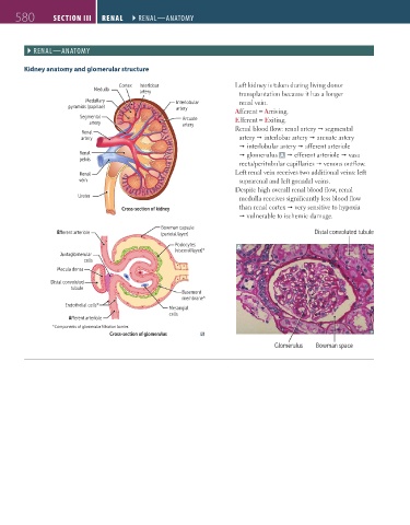
                                           Cortex  Interlobar                 Left kidney is taken during living donor
                                    Medulla
                                                 artery                        transplantation because it has a longer
                                 Medullary                  Interlobular       renal vein.
                            pyramids (papillae)             artery            Afferent = Arriving.
                               Segmental                      Arcuate         Efferent = Exiting.
                                  artery                      artery
                                Renal                                         Renal blood flow: renal artery Ž segmental
                                artery                                         artery Ž interlobar artery Ž arcuate artery
                                                                               Ž interlobular artery Ž afferent arteriole
                               Renal                                           Ž glomerulus  A  Ž efferent arteriole Ž vasa
                               pelvis
                                                                               recta/peritubular capillaries Ž venous outflow.
                               Renal                                          Left renal vein receives two additional veins: left
                               vein                                            suprarenal and left gonadal veins.
                                                                              Despite high overall renal blood flow, renal
                               Ureter                                          medulla receives significantly less blood flow
                                            Cross-section of kidney            than renal cortex Ž very sensitive to hypoxia
                                                                               Ž vulnerable to ischemic damage.
                                                        Bowman capsule
                         E erent arteriole              (parietal layer)                             Distal convoluted tubule
                                                            Podocytes         A
                                                            (visceral layer)*
                         Juxtaglomerular
                                 cells
                         Macula densa
                       Distal convoluted
                             tubule
                                                              Basement
                                                              membrane*
                           Endothelial cells*
                                                          Mesangial
                                                          cells
                            A erent arteriole
                       *Components of glomerular filtration barrier.
                                        Cross-section of glomerulus
                                                                                          Glomerulus  Bowman space


          FAS1_2019_14-Renal.indd   580                                                                                 11/7/19   5:42 PM
   619   620   621   622   623   624   625   626   627   628   629