
Page 663 - First Aid for the USMLE Step 1 2020, Thirtieth edition [MedicalBooksVN.com]_Neat
P. 663
RepRoductive ` REPRODUCTIVE—EmbRyOlOgy RepRoductive ` REPRODUCTIVE—EmbRyOlOgy SectioN iii 619
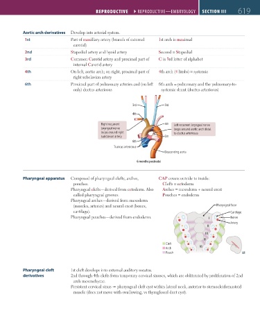
Aortic arch derivatives Develop into arterial system.
1st Part of maxillary artery (branch of external 1st arch is maximal
carotid)
2nd Stapedial artery and hyoid artery Second = Stapedial
3rd Common Carotid artery and proximal part of C is 3rd letter of alphabet
internal Carotid artery
4th On left, aortic arch; on right, proximal part of 4th arch (4 limbs) = systemic
right subclavian artery
6th Proximal part of pulmonary arteries and (on left 6th arch = pulmonary and the pulmonary-to-
only) ductus arteriosus systemic shunt (ductus arteriosus)
3rd 3rd
4th
Right recurrent 4th Left recurrent laryngeal nerve
laryngeal nerve loops around aortic arch distal
loops around right to ductus arteriosus
subclavian artery 6th
6th
Truncus arteriosus
Descending aorta
6 months postnatal
Pharyngeal apparatus Composed of pharyngeal clefts, arches, CAP covers outside to inside:
pouches. Clefts = ectoderm
Pharyngeal clefts—derived from ectoderm. Also Arches = mesoderm + neural crest
called pharyngeal grooves. Pouches = endoderm
Pharyngeal arches—derived from mesoderm
(muscles, arteries) and neural crest (bones, Pharyngeal floor
cartilage). Cartilage
Pharyngeal pouches—derived from endoderm. I Nerve
Artery
II
III
IV
Cleft
Arch VI
Pouch
Pharyngeal cleft 1st cleft develops into external auditory meatus.
derivatives 2nd through 4th clefts form temporary cervical sinuses, which are obliterated by proliferation of 2nd
arch mesenchyme.
Persistent cervical sinus pharyngeal cleft cyst within lateral neck, anterior to sternocleidomastoid
muscle (does not move with swallowing, vs thyroglossal duct cyst).
FAS1_2019_15-Repro.indd 619 11/7/19 5:52 PM