
Page 171 - fbkCardioDiabetes_2017
P. 171
Mechanism, Clinical Presentation and 147
Treatment of Diabetic Kidney Diseases
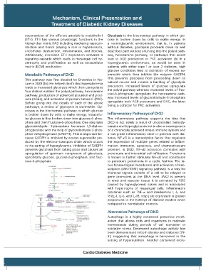
soconstrictor of the efferent arteriole is endothelin-1 Glycolysis is the biochemical pathway in which glu-
(ET-1). ET-1 has various physiologic functions in the cose is broken down by cells to make energy. In
kidney that mimic RAS including mediating vasocon- a normoglycemic environment, that is, in patients
striction and hence playing a role in hypertension, without diabetes, glycolysis proceeds down its well
endothelial dysfunction, inflammation, and fibrosis. described path without shunting into the polyol path-
Additionally, increased ET-1 expression activates a way, hexosamine pathway, or pathways that would
signaling cascade which leads to mesangial cell hy- lead to AGE production or PKC activation. (b) In a
pertrophy and proliferation as well as extracellular hyperglycemic environment, as would be seen in
matrix (ECM) production. patients with either type 1 or type 2 diabetes, high
glucose conditions lead to activation of excess su-
Metabolic Pathways of DKD peroxide which then inhibits the enzyme GADPH.
This prevents glycolysis from proceeding down its
This pathway was first detailed by Brownlee in Na-
ture in 2001 [16]. He helped clarify that hyperglycemia natural course and creates a backlog of glycolysis
leads to increased glycolysis which then upregulates precursors. Increased levels of glucose upregulate
four distinct entities: the polyol pathway, hexosamine the polyol pathway whereas increased levels of fruc-
pathway, production of advanced glycation end prod- tose-6-phosphate upregulate the hexosamine path-
ucts (AGEs), and activation of protein kinase C (PKC). way. Increased levels of glyceraldehyde-3-phosphate
Before going into the details of each of the above upregulate both AGE precursors and DAG, the latter
pathways, a review of glycolysis is worthwhile. Gly- being a cofactor for PKC activation.
colysis is the biochemical pathway in which glucose
is broken down by cells to make energy. Intracellu- Inflammatory Pathways of DKD
lar glucose is first broken down into glucose-6-phos- The inflammatory pathway supports the idea that
phate and then fructose-6-phosphate. One step later DKD is not solely a result of uncontrolled hemody-
glyceraldehyde- 3-phosphate becomes 1,3-diphos- namics and hyperglycemia but is also a consequence
phoglycerate with the help of glyceraldehyde-3-phos- of a chronically activated innate immune system and
phate dehydrogenase (GADPH) . This is important be- a low-grade inflammatory state in patients with dia-
cause GADPH is inhibited by excess superoxide pro- betes. NF-κB is a transcription factor that regulates
duced by the electron-transport chain which occurs the expression of multiple genes related to inflam-
in the setting of hyperglycemia. Inhibition of GADPH mation, immunity, apoptosis, and chemoattractant
prevents glycolysis from taking place and causes an protein-1. In DKD, NF-κB activation correlates with
upregulation of upstream component of glycolysis, proteinuria and interstitial cell infiltration. Proteinuria
specifically glucose, glucose-6-phosphate, and fruc- is known to further stimulate NF-κB and contributes
tose-6-phosphate to persistent proteinuria in a cyclic fashion. The Ja-
nus kinase/signal transducers and activators of tran-
scription (JAK/STAT) signaling pathway is a way for
chemical signals outside of a cell to be relayed to
gene promoters at the DNA level. JAK2 is present
in renal and vascular tissue. It is activated by ROS
caused by hyperglycemic states and is associated
with hypertrophy of mesangial cells. Inflammatory
cytokines such as TNF-κ and interleukins 1, 6, and
18 (IL-1, IL-6, and IL-18, resp.) are expressed in greater
proportions in the kidneys of diabetic models when
compared to nondiabetic controls.
Alternative Pathways of DKD
Autophagy is a highly conserved protective mech-
anism that allows cells and organisms to maintain
homeostasis during periods of cell starvation or
oxidative stress. Decreased autophagic activity has
been demonstrated in both obesity and diabetes [29-
. 31] suggesting that autophagy is hampered in the
setting of hypernutrition Another conserved evolu-
Cardio Diabetes Medicine