
Page 249 - fbkCardioDiabetes_2017
P. 249
Bradyarrhythmias 225
Second degree AV block: Risk stratification
Diagnosis: Type 1:
Mobitz type 1: progressive lengthening of the PR in- • Disease is at the AV node
terval until one P wave is totally blocked and produc- • Usually benign and self-limiting
es no QRS (fig 10).The associated QRS is typically • Unlikely to progress to complete heart block
of normal duration. This is usually due to AV node • Usually associated with inferior wall MI
fatigue. After a pause during which the AV node re-
covers, this cycle is repeated. This is called Wencke- Type 2:
bach’s phenomenon. • Block occurs at the distal conducting system
• Indicates severe disease
• Progresses to complete heart block; patient may
have Stoke – Adams attacks because escape
rhythm is from the ventricle
• Requires active treatment including pacemaker
Fig 10: progressive lengthening of PR interval followed 2: 1 AV block
by a blocked P wave When every other P wave is not conducted to the
ventricles, it is 2: 1 AV block. The block can be in
Mobitz type 2: PR interval in the conducted beats is
fixed but there is a sudden non conduction of a P the AV node or in the HIS Purkinje system. Clue to
wave in the presence of constant and consecutive the site of block can be obtained by looking at the
conduction width of the QRS complex. (if the block is in the HIS
Purkinje syst em, QRS is wide)
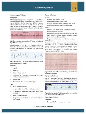
Fig 11: Mobitz type 2 AV block where there is a sudden
non conduction of P wave
Etiology
• Type 1
• Athletes
Figure 12: 2:1 AV block demonstrating non conduction
• Acute inferior wall MI
nd
of every 2 P wave
• Drugs like beta blockers, digoxin, calcium chan- High grade AV block:
nel blockers, amiodarone
The term high grade AV block is applied to a pattern
• Myocarditis where ≥ 2 sinus P waves are blocked consecutively in
• Post mitral valve surgery, TOF repair the context of periodic AV conduction (fig 13).
Type 2
• Acute anterior wall MI
• Idiopathic fibrosis of the conducting system
• Inflammatory conditions (rheumatic fever, myo-
carditis) Figure 13: high grade AV block showing non conduc-
tion of > 2 consecutive P waves
• Autoimmune diseases
Third degree AV block
• Drugs like beta blockers, calcium channel block-
ers, digoxin Diagnosis
• None of the P waves are conducted
Cardio Diabetes Medicine