
Page 247 - fbkCardioDiabetes_2017
P. 247
Bradyarrhythmias 223
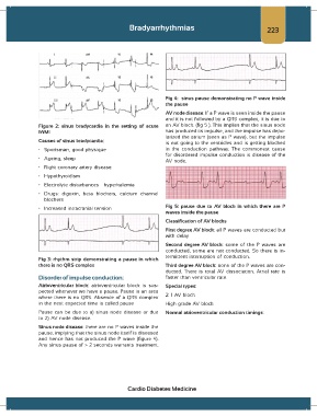
Fig 4: sinus pause demonstrating no P wave inside
the pause
AV node disease: If a P wave is seen inside the pause
and it is not followed by a QRS complex, it is due to
Figure 2: sinus bradycardia in the setting of acute an AV block. (fig 5 ). This implies that the sinus node
IWMI has produced its impulse, and the impulse has depo-
larized the atrium (seen as P wave), but the impulse
Causes of sinus bradycardia: is not going to the ventricles and is getting blocked
• Sportsman, good physique in the conduction pathway. The commonest cause
for disordered impulse conduction is disease of the
• Ageing, sleep
AV node.
• Right coronary artery disease
• Hypothyroidism
• Electrolyte disturbances – hyperkalemia
• Drugs: digoxin, beta blockers, calcium channel
blockers
• Increased intracranial tension Fig 5: pause due to AV block in which there are P
waves inside the pause
Classification of AV blocks
First degree AV block: all P waves are conducted but
with delay
Second degree AV block: some of the P waves are
conducted, some are not conducted. So there is in-
termittent interruption of conduction.
Fig 3: rhythm strip demonstrating a pause in which
there is no QRS complex Third degree AV block: none of the P waves are con-
ducted. There is total AV dissociation. Atrial rate is
Disorder of impulse conduction: faster than ventricular rate.
Atrioventricular block: atrioventricular block is sus- Special types:
pected whenever we have a pause. Pause is an area
where there is no QRS. Absence of a QRS complex 2: 1 AV block
in the next expected time is called pause High grade AV block
Pause can be due to a) sinus node disease or due Normal atrioventricular conduction timings:
to 2) AV node disease.
Sinus node disease: there are no P waves inside the
pause, implying that the sinus node itself is diseased
and hence has not produced the P wave (figure 4).
Any sinus pause of > 2 seconds warrants treatment.
Cardio Diabetes Medicine