
Page 359 - fbkCardioDiabetes_2017
P. 359
Cardio Diabetes Medicine 2017 335
(PSD) and Phase Histogram Bandwidth al flow reserve estimation have been found to have
(PHB). Mukherjee et al. in a study of non good prognostic value. Normal perfusion on SPECT
ischemic dilated cardiomyopathy patients or myocardial flow reserve on PET predicts an excel-
lent prognosis in the intermediate term (2–5 years).
noted that responders had significantly
higher dyssynchrony indices of PSD (64 ± Prognosis and risk stratification in heart
17° vs 39 ± 13°; P<.01) and PHB (215 ± 64° vs failure:
110 ± 44°; P<.01) compared to nonrespond- Patients with HF show increased activation of the
ers. ROC curve analysis demonstrated that sympathetic nervous system, reflected by an increase
the maximum accuracy for prediction of CRT in plasma norepinephrine levels. In addition, neuronal
response was obtained with values of 128° uptake of norepinephrine is impaired in the failing
for PHB and 43° for PSD (86% sensitivity and myocardium. Both the enhanced release of norepi-
nephrine and changes in its cardiac neuronal uptake
80% specificity for both parameters) may be responsible for the observed downregulation
(Figure 1) [5] of adrenoreceptors in patients with HF [6]. Myocar-
dial innervation imaging with I-123 meta-iodo-benzyl-
guanidine (MIBG) scintigraphy provides a noninvasive
tool for the investigation of cardiac sympathetic in-
nervation. Increased norepinephrine turnover and
pre-synaptic norepinephrine deficits can be identified
as an increased MIBG washout rate (WR) from the
heart and decreased MIBG activity quantified as the
heart-to mediastinum (H/M) ratio. While the early and
delayed H/M ratios and the wash rate are thought
to reflect specific aspects of the MIBG uptake, stor-
age and release mechanisms, the delayed H/M ra-
tio has been found to be the strongest predictor of
HF prognosis in clinical studies. ADMIRE-HF study
revealed that patients with delayed H/M ratio > 1.6
had significantly better prognosis when compared
with patients with H/M ratio < 1.6 [7]. Assessment
of cardiac dyssynchrony has also been used in risk
stratification and prognostication of non-ischemic
cardiomyopathy patients [8].
Molecular imaging of HF
Molecular mechanisms of HF are operative at the
preclinical stage and imaging these mechanisms
may lead to understanding of HF pathophysiology,
and starting early therapy to halt disease progres-
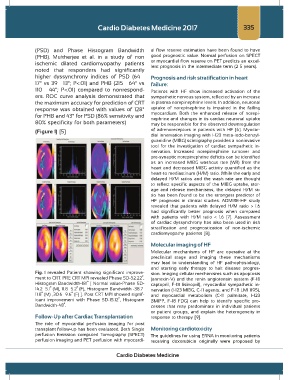
Fig. 1 revealed Patient showing significant improve- sion. Imaging cellular mechanisms such as apoptosis
ment to CRT. PRE CRT MPI revealed Phase SD-52.22˚, (annexin-V) and the renin angiotensin system (F-18
Histogram Bandwidth-181˚ [ Normal value-Phase SD- captopril, F-18 lisinopril), myocardial sympathetic in-
14.2± 5.1˚ (M), 11.8± 5.2˚ (F), Histogram Bandwidth -38.7± nervation (I-123 MIBG, C-11 agents, and F-18 LMI 1195),
11.8˚ (M) ,30.6± 9.6˚ (F) ]. Post CRT MPI showed signif- and myocardial metabolism (C-11 palmitate, I-123
icant improvement with Phase SD-15.12˚, Histogram BMIPP, F-18 FDG) can help to identify specific pro-
Bandwidth-48˚. cesses that may predominate in individual patients
or patient groups, and explain the heterogeneity in
Follow-Up after Cardiac Transplantation response to therapy [9].
The role of myocardial perfusion imaging for post
transplant follow-up has been evaluated. Both Single Monitoring cardiotoxicity
perfusion Emission computed Tomography (SPECT) The guidelines for using ERNA in monitoring patients
perfusion imaging and PET perfusion with myocardi- receiving doxorubicin originally were proposed by
Cardio Diabetes Medicine