
Page 360 - fbkCardioDiabetes_2017
P. 360
Role of Nuclear Imaging in The Evaluvation of Non Coronary
336
Artery Disease
Schwartz and co-workers [10] and have been adopt- potential to improve the early detection of myocardial
ed widely. These guidelines indicate a baseline LVEF inflammation, enable quantification of disease activi-
evaluation before beginning chemotherapy or before ty, guide therapeutic interventions, and monitor treat-
100 mg/m2 doxorubicin administration. Further serial ment success. Leukocyte scintigraphy is highly spe-
follow-up studies are based on the patient cardiac cific for infection because granulocytes are recruited
function, risk factors, and doxorubicin dose. If the to the site of infection. Whereas general utility has
baseline calculated LVEF is ≤ 30%, doxorubicin should been compromised by limited sensitivity, the imple-
not be chosen as an agent for therapy; if LVEF is mentation of single photon emission computed to-
from >30% to < 50%, follow-up ERNA should be ob- mography (SPECT) imaging has increased diagnostic
tained before each dose and doxorubicin therapy dis- performance and opened new possibilities in settings
continued when LVEF declines ≥ to 10% and/or LVEF in which high specificity is needed, e.g., in endocardi-
is ≤ 30%; if LVEF is ≥50%, ERNA should be obtained at tis imaging. Since increased glucose metabolism due
dose of 250-300 mg/m2, 400-450 mg/m2 (at 400 to overexpression of glucose transporters and over-
mg/m2 in patient with risk factors), and before each production of glycolytic enzymes in inflammatory
higher doses after. Doxorubicin should be discontin- cells considered as a hallmark of inflammation FDG
ued if the LVEF decreases to ≥ 10% to a level of ≤ 50%. PET-CT is the standard of reference for molecular im-
A review article by Mitra et al. describes various utility aging of myocardial inflammation. Indications include
of MUGA stuy in great details [11] endocarditis, myocarditis, or sarcoidosis but as well
the detection of inflammatory changes after acute
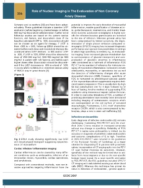
(Figure 2)
myocardial infarction (AMI). However, specificity of
FDG is hampered by physiological glucose uptake
of the myocardium whose suppression requires dedi-
cated patient preparation. This is usually done by high
fat low carbohydrate diet for 3 days followed by 12
hour of fasting. Another method of suppressing FDG
uptake is using intravenous heparin before the scan.
In order to overcome limitations of FDG, a number of
promising alternatives have recently been introduced
including imaging of somatostatin receptors which
are overexpressed on the cell surface of activated
macrophages. Furthermore, C-X-C motif chemokine
receptor CXCR4, which is also overexpressed by leu-
kocytes, plays a role in stem cell trafficking [12].
Infective endocarditis:
Early diagnosis of infective endocarditis (IE) remains
challenging. Combining FDG-PET/CT with the mod-
ified Duke Criteria resulted in increased sensitivity
without any change in specificity. Reliability of FDG
PET-CT in native valve endocarditis is limited, but its
accuracy in diagnosis of prosthetic valve endocarditis
and systemic complications of IE is high. Therefore
Fig. 2 ERNA study showing significantly low LVEF in 2015 FDG-PET/CT was included in the guidelines
with broad phased histogram suggesting dysynchro- of the European Society of Cardiology as a major
nous LV myocardium criterion for diagnosing IE in patients with prosthetic
valves. Incorporation of CT angiography into the PET/
Cardiac Infection/ inflammation imaging:
CT scan further improves its sensitivity.. However,
Cardiac inflammation can be caused by many differ- specificity of the method may be limited due to arti-
ent conditions such as endocarditis, infection of an facts from metal implants or due to the non-specific
intracardiac device, myocarditis, cardiac sarcoidosis, biologic tracer signal. As a more specific alternative
and amyloidosis. to FDG-PET/CT, the ESC guidelines included SPECT/
CT imaging with radiolabeled autologous white blood
Compared with conventional methods, new non-in-
vasive approaches targeting inflammation have the cells (WBC). Whereas this technique has proven its
value in detection of endocarditis , general applica-
GCDC 2017