
Page 399 - fbkCardioDiabetes_2017
P. 399
Cardio Diabetes Medicine 2017 375
Conclusion :
Hybrid PET/MR imaging could also become the
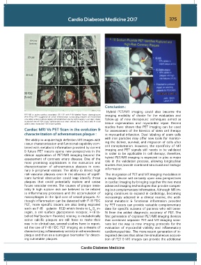
PET MR in active cardiac sarcoidosis 3D LGE with F-18 labelled fluoro- deoxyglucose imaging modality of choice for the evaluation and
(F-18 FDG) PET suggestive of active inflammation surrounding requires of established
scar white arrows indicate regions of established scar by LGE imaging . Last rows shows follow-up of new therapeutic techniques aimed at
increased row of FDG tracer uptake entrance rows around the scar areas with & small
spleen rows increased FDG tracer uptake. tissue regeneration and myocardial repair. Recent
studies have shown that PET imaging can be used
Cardiac MRI Vs PET Scan in the evolution & for assessment of the kinetics of stem cell therapy
characterization of atheramatous plaque : in myocardial infarction. Dual labeling of stem cells
The ability to acquire high definition MR images with with iron particles may offer new tools for monitor-
tissue characterization and functional capability com- ing the deliver, survival, and migration of cells after
bined with metabolic information provided by current cell transplantation. However, the specificity of MR
& future PET tracers opens new perspectives in the imaging and PET signals still needs to be validated
clinical application of PET/MR imaging beyond the in order to be applicable in cell therapy; therefore,
assessment of coronary artery disease. One of the hybrid PET/MR imaging is expected to play a major
most promising applications is the evaluation and role in the validation process, allowing longitudinal
characterization of atheramatous plaques in coro- studies that provide combined structural and biologic
nary & peripheral vassals. The ability to detect high information.
risk vascular plaques even in the absence of signif- The integration of PET and MR imaging modalities in
icant luminal obstruction could leap identify those a single device will certainly open new perspectives
plaques that could potentially rupture and cause in cardiac imaging by bringing together the two most
future vascular events. The causes of plaque insta- advanced imaging technologies that provide compet-
bility & high rupture risk are believed to be related ing but complementary information. Although MR im-
to inflammatory processes and the accumulation of aging continues to expand in cardiology, becoming
macrophages in the lipid core of vessel plaques. Al- increasingly adopted in a clinical routine, the addi-
though inflammation can be detected with F-18 FDG tional metabolic & functional information provided
PET, more specific tracers are also being explored by PET tracers can provide valuable complementary
such as F-18 – galacto –RGD which binds to L B in- data for specific subsets of patients who will bene-
v
3
tegrin, a cell surface glycoprotein receptor, F-18 la- fit from the added diagnostic accuracy of PET. The
belled NaF(sodium fluoride) relating to metabolically first generation of coplanar PET/MR imaging devices
active calcific plaques are still have to make their that combined separate PET and MR imaging scan-
way in to clinical use, several studies have to report- ners led the way to new imaging protocols for the
ed the use of F-18 FDG PET imaging as a means of evaluation of myocardial viability and inflammatory
characterizing inflammatory activity in atherosclerotic cardiomyopathies . The more recent generation of in-
plaque and thus as a surrogate biomarker for detect- tegrated devices that allow the simultaneous acquisi-
ing vulnerable plaques. tion of PET & MR images can provide the additional
Cardio Diabetes Medicine