
Page 396 - fbkCardioDiabetes_2017
P. 396
372 Cardiac MRI vs PET Scan
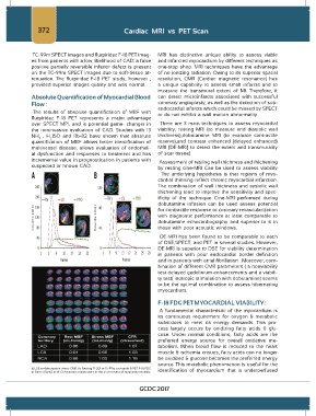
TC-99m SPECT images and flurpiridaz F-18 PET imag- MRI has distinctive unique ability to assess viable
es from patients with a low likelihood of CAD: a false and infarcted myocardium by different techniques as
positive partially reversible inferior defect is present one-stop shop. MRI techniques have the advantage
on the TC-99m SPECT images due to soft-tissue at- of no ionizing radiation. Owing to its superior spatial
tenuation. The flurpiridaz F-18 PET study, however , resolution, CMR (Cardiac magnetic resonance) has
provided superior images quality and was normal. a unique capability to assess small infarcts and to
measure the transmural extent of MI. Therefore, it
Absolute Quantification of Myocardial Blood can detect microinfarcts associated with successful
Flow : coronary angioplasty, as well as the detection of sub-
endocardial infarcts which could be missed by SPECT
The results of absolute quantification of MBF with or do not exhibit a wall motion abnormality.
flurpiridaz F-18 PET represents a major advantage
over SPECT MPI, and is potential game- changer in There are 3 main techniques to assess myocardial
the noninvasive evaluation of CAD. Studies with 13 viability; resting MRI (to measure end diastolic wall
NH , H 15O and Rb-82 have shown that absolute thickness),dobutamine MRI (to evaluate contractile
3
2
quantification of MBF allows better identification of reserve),and contrast enhanced (delayed enhanced)
multivessel disease, allows evaluation of endotheli- MRI [DE-MRI] to detect the extent and transmurality
al dysfunction and responses to treatment and has of scar tissue)
incremental value in prognostication in patients with Assessment of resting wall thickness and thickening
suspected or known CAD.
by resting cine-MRI Can be used to assess viability
. The underlying hypothesis is that regions of myo-
cardial thinning reflect chronic myocardial infarction.
The combination of wall thickness and systolic wall
thickening tend to improve the sensitivity and spec-
ificity of the technique. Cine-MRI performed during
dobutamine infusion can be used assess potential
for contractile response to coronary revascularization
with diagnostic performance at least comparable to
dobutamine echocardiography and superior to it in
those with poor acoustic windows.
DE-MRI has been found to be comparable to each
of DSE,SPECT, and PET in several studies. However,
DE-MRI is superior to DSE for viability determination
in patients with poor endocardial border definition
and in patients with atrial fibrillation . Moreover, com-
bination of different CMR parameters ( a nonviability
test delayed gadolinium enhancements and a viabili-
ty test( inotropic stimulation with dobutamine) seems
to be the optimal combination to assess hibernating
myocardium.
F-18 FDG PET MYOCARDIAL VIABILITY :
A fundamental characteristic of the myocardium is
its continuous requirement for oxygen & metabolic
substrates to meet its energy demands. This pro-
cess largely occurs by oxidizing fatty acids & glu-
cose. Under normal conditions, fatty acids are the
preferred energy source for overall oxidative me-
tabolism. When blood flow is reduced to the heart
muscle & ischemia ensues, fatty acids can no longer
be oxidized & glucose becomes the preferred energy
source. This metabolic phenomenon is useful for the
ii) LGE or dobutamine stress CMR Vs Resting Tl-201 or Tc 99m sestamibi & PET F-18 FDG identification of myocardium that is underperfused
or Stress Rb-82 or N-13 Ammonia radiotracers in the assessment of myocardial viability.
GCDC 2017