
Page 395 - fbkCardioDiabetes_2017
P. 395
Cardio Diabetes Medicine 2017 371
negative charge, 18FDG-6- phosphate is trapped in-
tracellularly.
The feasibility and accuracy of exercise 18FDG im-
aging for hot spot imaging of exercise induced
myocardial ischemia was studied by performing si-
multaneous exercise perfusion and 18FDG imaging
in patient scheduled to undergo exercise and rest
myocardial perfusion imaging for suspected CAD.
All patients underwent a symptom –limited exercise
after an overnight fast. 99mTc-sestamibi (25mci) and
18FDG (8-10mCi) were injected iv at peak exercise.
The patients underwent imaging using a dual-head Weishaowu
large field of view single –crystal SPECT imaging cam-
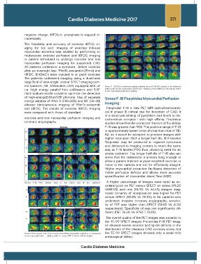
era (varicam ,GE ,Milwaukee,USA) equipped with ul- Stress F -18 FDG ischaemia imaging showing intense F-18 FDG uptake in the inferior &
tra –high energy parallel hole collimators and 5/8” lateral walls of the myocardium which was showing a fixed defect in the regular stress
& rest myocardial perfusion imaging.
thick sodium iodide crystal to optimize the detection
of high-energy(511KeV)18F photons. Use of separate Stress F-18 Flurpiridaz Myocardial Perfusion
energy window of 99m Tc (140+20%) and 18F (511+30)
allowed simultaneous imaging of 99mTc-sestamibi Imaging :
and 18FDG. The results of exercise 18FDG imaging Flurpiridaz F-18 a new PET MPI radiopharmaceuti-
were compared with those of standard cal in phase III clinical trial for detection of CAD. It
is a structural analog of pyridaben and binds to mi-
exercise and rest myocardial perfusion imaging and tochondrian complex I with high affinity. Preclinical
coronary angiography. studies show that the extraction fraction of flurpiridaz
F-18 was greater than 90%. The positron range of F-18
is approximately seven times shorter than that of RB-
82, so it would be accepted to produce images with
higher resolution. With a longer half-life, 18 F-labeled
flurpiridaz may be produced at regional cyclotrons
and delivered to imaging centers in much the same
way as F-18 labeled FDG thus ,obviating need for an
onsite cyclotron. The longer half-life of F-18 also en-
sures that the radiotracer is present long enough to
allow a patient injected at peak treadmill exercise to
move to the camera and still be effectively imaged.
Higher myocardial extraction facilitates detection of
milder perfusion defects and allows more accurate
quantification of myocardial blood flow (MBF).
A higher percentage of images were rated as ex-
Intense F-18 FDG uptake seen in the lateral wall of the segments
cellent/good on PET versus SPECT on stress (99.2%
Short Axis
Vs88.5%) and rest (96.9% Vs 66.4%) images diag-
Ex
nostic certainty of interpretation was higher for PET
R versus SPECT (90.8% Vs 70.9%). In 86 patients who
underwent invasive coronary angiography, sensitiv-
ity of PET was higher than SPECT (78.8% Vs 61.5%
Vertical Long Axis Horizontal Long Axis respectively). Specificity of was not significantly dif-
Ex ferent (PET 76.5% Vs SPECT 73.5%).
The overall quality of the PET images was superior to
R
the TC-99 SPECT images. Flurpiridaz F-18 PET imag-
es showed severe anterior and apical defects in the
distribution of the diseases CAD coronary artery, but
gx Jain et al
intense FDG uptake seen in the lateral and inferior myocardial segments as compared the TC-99 SPECT images showed only a small mild
to no stress perfusion defects seen in stress MPI in the Ist row of images. anteroapical defect.
Cardio Diabetes Medicine