Page 398 - fbkCardioDiabetes_2017
P. 398

374                                Cardiac  MRI  vs  PET Scan


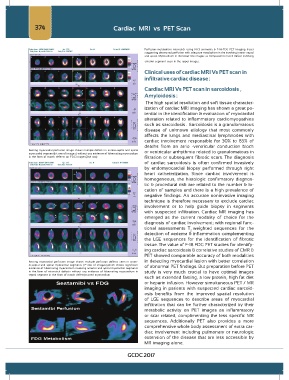
                                                                 Perfusion-metabolism  mismatch  using N-13  ammonia  &  F-18-FDG PET imaging  tracer
                                                                 suggesting decreased perfusion with adequate metabolism in the involving intero- septal
                                                                 and apical. Myocardium in the lower row images as compared to match defect involving
                                                                 anterior segment seen in the upper images.

                                                                 Clinical uses of cardiac MRI Vs PET scan in
                                                                 infiltrative cardiac disease :

                                                                 Cardiac MRI Vs PET scan in sarcoidosis ,
                                                                 Amyloidosis :

                                                                  The high spatial resolution and soft tissue character-
                                                                 ization of cardiac MRI imaging has shown a great po-
                                                                 tential in the identification & evaluation of myocardial
                                                                 alteration  related to inflammatory  cardiomyopathies
                                                                 such as sarcoidosis . Sarcoidosis is a granulomatous
                                                                 disease  of unknown  etiology  that  most commonly
                                                                 affects  the lungs and  mediastinal lymphnodes with
                                                                 cardiac involvement  responsible  for  30% to 85% of
                                                                 deaths  from an atrio -ventricular conduction  block
              Resting myocardial perfusion image shows multiple defects in antero-septal and apical   or ventricular arrhythmia related to granulomatous in
              myocardial segment(Ist row of images.) without any evidence of hibernating myocardium
              in the form of match defects on FDG images(2nd row)  filtration or subsequent fibrotic scars. The diagnosis
                                                                 of  cardiac sarcoidosis  is  often confirmed invasively
                                                                 by  endomyocardial  biopsy  performed  through right
                                                                 heart catheterization. Since  cardiac involvement is
                                                                 homogeneous, the  histologic confirmatory  diagnos-
                                                                 tic & procedural risk are related to the number & lo-
                                                                 cation of samples and there is a high prevalence of
                                                                 negative findings.  An accurate noninvasive  imaging
                                                                 technique is  therefore  necessary  to exclude cardiac
                                                                 involvement  or  to help  guide  biopsy  in segments
                                                                 with suspected  infiltration. Cardiac MR imaging  has
                                                                 emerged  as the current modality of choice  for  the
                                                                 diagnosis of cardiac involvement; with regional func-
                                                                 tional  assessments T weighted sequences for the
                                                                                     2
                                                                 detection of oedema & inflammation complementing
                                                                 the  LGE  sequences for the  identification  of fibrotic
                                                                 tissue. The value of F-18 FDG PET studies for identify-
                                                                 ing cardiac sarcoidosis & correlative studies of CMR &
                                                                 PET showed comparable accuracy of both modalities
              Resting myocardial perfusion  image  shows multiple perfusion  defects seen  in anteri-  in detecting myocardial lesion with better correlation
              or,septal  and  apical  myocardial  segments  (1 st   row  of  images),which  shows  significant   of abnormal PET findings. But preparation before PET
              evidence of hibernating myocardium involving anterior and apical myocardial segments
              in the form of mismatch defects without  any evidence of hibernating myocardium  in   study is  very  much  crucial  to have  optimal images
              septal segment in the form of match defect(scarred myocardium
                                                                 such as extended fasting, a low protein, high fat diet
                                                                 or heparin infusion. However simultaneous PET / MR
                                                                 imaging in patients with  suspected cardiac  sarcoid-
                                                                 osis  benefits from the improved  spatial  resolution
                                                                 of LGE sequences  to describe  areas  of myocardial
                                                                 infiltration that  can  be further characterized  by their
                                                                 metabolic activity on PET images  as inflammatory
                                                                 or  scar  related,  complimenting  the less  specific  MR
                                                                 sequences.  Additionally  PET  also  provides  a more
                                                                 comprehensive whole body assessment of extra car-
                                                                 diac  involvement  including  pulmonary or neurologic
                                                                 extension of the disease that are less accessible by
                                                                 MR imaging alone.

                                                         GCDC 2017
   393   394   395   396   397   398   399   400   401   402   403