
Page 102 - Critical Care Nursing Demystified
P. 102
Chapter 3 CARE OF THE PATIENT WITH CRITICAL CARDIAC AND VASCULAR NEEDS 87
Aortic arch
Pulmonary artery
Superior Aortic valve
vena cava
Left pulmonary
veins
Right
pulmonary
veins Left atrium
Pulmonic Mitral valve
valve
Chordae
Right atrium tendinae
Papillary
muscles
Tricuspid
valve Left ventricle
Right
ventricle
Inferior Downloaded by [ Faculty of Nursing, Chiangmai University 5.62.158.117] at [07/18/16]. Copyright © McGraw-Hill Global Education Holdings, LLC. Not to be redistributed or modified in any way without permission.
vena cava
Unoxygenated blood (gray)
Oxygenated blood (red) Parietal
pericardium
Endocardium
Pericardial
Myocardium space
Epicardium
Visceral
pericardium
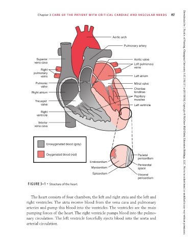
FIGURE 3–1 • Structure of the heart.
The heart consists of four chambers, the left and right atria and the left and
right ventricles. The atria receive blood from the vena cava and pulmonary
arteries and pump this blood into the ventricles. The ventricles are the main
pumping forces of the heart. The right ventricle pumps blood into the pulmo-
nary circulation. The left ventricle forcefully ejects blood into the aorta and
arterial circulation.