
Page 107 - Critical Care Nursing Demystified
P. 107
92 CRITICAL CARE NURSING DeMYSTIFIED
An abnormal pulsation that can be seen on inspection of the neck is jugular
venous distention (JVD). With the patient lying supine and the head elevated
30 to 45 degrees, you should not see visible pulsation at the side of the neck.
JVD is a response to increased intrathoracic pressure during the Valsalva
maneuver and can temporarily be seen normally when a weightlifter bears
down while lifting weights. If you see visible pulsations that occur above the
jaw line, this might indicate an increase in circulating volume to the right side
of the heart, which can be caused by right-sided heart failure.
NURSING ALERT
Inspect the jugular veins for pulsations and distention, which might be apparent and
indicative of right-sided cardiac failure.
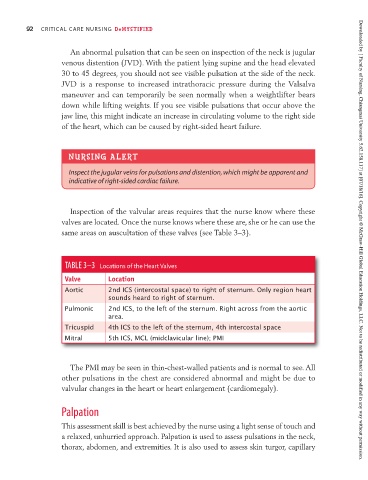
Inspection of the valvular areas requires that the nurse know where these
valves are located. Once the nurse knows where these are, she or he can use the
same areas on auscultation of these valves (see Table 3–3). Downloaded by [ Faculty of Nursing, Chiangmai University 5.62.158.117] at [07/18/16]. Copyright © McGraw-Hill Global Education Holdings, LLC. Not to be redistributed or modified in any way without permission.
TABLE 3–3 Locations of the Heart Valves
Valve Location
Aortic 2nd ICS (intercostal space) to right of sternum. Only region heart
sounds heard to right of sternum.
Pulmonic 2nd ICS, to the left of the sternum. Right across from the aortic
area.
Tricuspid 4th ICS to the left of the sternum, 4th intercostal space
Mitral 5th ICS, MCL (midclavicular line); PMI
The PMI may be seen in thin-chest-walled patients and is normal to see. All
other pulsations in the chest are considered abnormal and might be due to
valvular changes in the heart or heart enlargement (cardiomegaly).
Palpation
This assessment skill is best achieved by the nurse using a light sense of touch and
a relaxed, unhurried approach. Palpation is used to assess pulsations in the neck,
thorax, abdomen, and extremities. It is also used to assess skin turgor, capillary