
Page 103 - Critical Care Nursing Demystified
P. 103
88 CRITICAL CARE NURSING DeMYSTIFIED
Pulmonary circulation begins at the pulmonary artery, which receives
venous blood from the right ventricle. The pulmonary artery divides into the
left and right main stem branches. Oxygenated blood returns to the left side
of the heart through the pulmonary veins.
Four cardiac valves exist to allow blood to flow in only one direction. The
two AV (atrioventricular valves) prevent the backflow of blood into the atria
during ventricular contraction. These AV valves are the tricuspid and mitral
valves. The semilunar valves, or the pulmonic and aortic valves, open during
systole, allowing blood to flow out of the ventricles. These valves will then close
to prevent blood regurgitation back into the ventricles.
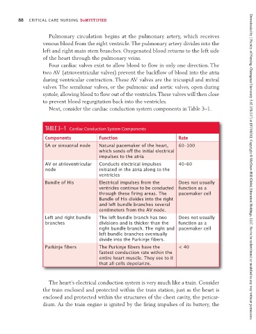
Next, consider the cardiac conduction system components in Table 3–1.
TABLE 3–1 Cardiac Conduction System Components
Components Function Rate
SA or sinoatrial node Natural pacemaker of the heart, 60–100
which sends off the initial electrical
impulses to the atria
AV or atrioventricular Conducts electrical impulses 40–60
node initiated in the atria along to the Downloaded by [ Faculty of Nursing, Chiangmai University 5.62.158.117] at [07/18/16]. Copyright © McGraw-Hill Global Education Holdings, LLC. Not to be redistributed or modified in any way without permission.
ventricles
Bundle of His Electrical impulses from the Does not usually
ventricles continue to be conducted function as a
through these firing areas. The pacemaker cell
Bundle of His divides into the right
and left bundle branches several
centimeters from the AV node.
Left and right bundle The left bundle branch has two Does not usually
branches divisions and is thicker than the function as a
right bundle branch. The right and pacemaker cell
left bundle branches eventually
divide into the Purkinje fibers.
Purkinje fibers The Purkinje fibers have the < 40
fastest conduction rate within the
entire heart muscle. They see to it
that all cells depolarize.
The heart’s electrical conduction system is very much like a train. Consider
the train enclosed and protected within the train station, just as the heart is
enclosed and protected within the structures of the chest cavity, the pericar-
dium. As the train engine is ignited by the firing impulses of its battery, the