
Page 104 - Critical Care Nursing Demystified
P. 104
Chapter 3 CARE OF THE PATIENT WITH CRITICAL CARDIAC AND VASCULAR NEEDS 89
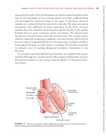
sinoatrial (SA) node of the heart transmits its initial electrical impulses to the
atria. As the train begins to move and gain speed, it is further conducted along
and encouraged by electrical charges to its engine. In the heart, electrical
impulses are conducted from the atria to the ventricles. The train increases its
momentum with additional electrical conduction. In the heart, electrical
impulses from the ventricles travel along through the Bundle of His and the
Purkinje fibers to create ventricular systole and diastole. The railroad tracks
diverge into many directions, as does the vascular system. The vascular system,
which is composed of numerous capillaries, veins and arteries, will receive its
precious cargo of oxygenated blood from the pulmonary circulation and direct
it throughout the body or to the systemic circulation. The train has completed
its intricate route of reaching designated circulatory destinations as has
the heart.
It is crucial to remember that the heart structures are supplied with oxygen-
ated blood through the coronary arteries. This oxygenated blood then travels to
the general circulation via the coronary veins. See Figure 3–2, Nervous Conduc-
tion System. Downloaded by [ Faculty of Nursing, Chiangmai University 5.62.158.117] at [07/18/16]. Copyright © McGraw-Hill Global Education Holdings, LLC. Not to be redistributed or modified in any way without permission.
Sinoatrial (SA) LA
node Ventricular
AV junction RA myocardium
AV node
His bundle
LV
Purkinje
fibers
RV
Left bundle
branch
Right bundle branch
Ventricular septum
FIGURE 3–2 • Nervous Conduction System. (Reproduced, with permission, from Fauci AS et al.
Harrison’s Principles of Internal Medicine, 17th ed. McGraw-Hill, 2008.)