
Page 142 - Critical Care Nursing Demystified
P. 142
Chapter 3 CARE OF THE PATIENT WITH CRITICAL CARDIAC AND VASCULAR NEEDS 127
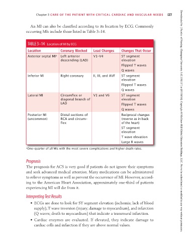
An MI can also be classified according to its location by ECG. Commonly
occurring MIs include those listed in Table 3–14.
TABLE 3–14 Location of MI by ECG
Location Coronary Blocked Lead Changes Changes That Occur
Anterior septal MI a Left anterior V1–V4 ST segment
descending (LAD) elevation
Flipped T waves
Q waves
Inferior MI Right coronary II, III, and AVF ST segment
elevation
Flipped T waves
Q waves
Lateral MI Circumflex or V1 and V6 ST segment
diagonal branch of elevation
LAD Flipped T waves
Q waves
Posterior MI Distal sections of Reciprocal changes
(uncommon) RCA and circum- (reverse as in back
flex of the heart) Downloaded by [ Faculty of Nursing, Chiangmai University 5.62.158.117] at [07/18/16]. Copyright © McGraw-Hill Global Education Holdings, LLC. Not to be redistributed or modified in any way without permission.
ST segment
elevation
T wave elevation
Large R waves
a One-quarter of all MIs with the most severe complications and higher death rates.
Prognosis
The prognosis for ACS is very good if patients do not ignore their symptoms
and seek advanced medical attention. Many medications can be administered
to relieve symptoms as well as prevent the occurrence of MI. However, accord-
ing to the American Heart Association, approximately one-third of patients
experiencing MI will die from it.
Interpreting Test Results
• ECGs are done to look for ST segment elevation (ischemia; lack of blood
supply), T wave inversion (injury; damage to myocardium), and infarction
(Q waves; death to myocardium) that indicate a transmural infarction.
• Cardiac enzymes are evaluated. If elevated, they indicate damage to
cardiac cells and infarction if they are above normal values.