
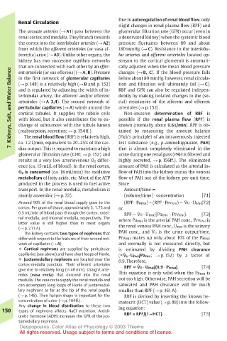
Page 163 - Color_Atlas_of_Physiology_5th_Ed._-_A._Despopoulos_2003
P. 163
Renal Circulation               Due to autoregulation of renal blood flow, only
                                       slight changes in renal plasma flow (RPF) and
       The arcuate arteries (! A1) pass between the  glomerular filtration rate (GFR) occur (even in
       renal cortex and medulla. They branch towards  a denervated kidney) when the systemic blood
       the cortex into the interlobular arteries (! A2)  pressure fluctuates between 80 and about
       from which the afferent arterioles (or vasa af-  180 mmHg (! C). Resistance in the interlobu-
       ferentia) arise (! A3). Unlike other organs, the  lar arteries and afferent arterioles located up-
       kidney has two successive capillary networks  stream to the cortical glomeruli is automati-
       that are connected with each other by an effer-  cally adjusted when the mean blood pressure
    Kidneys, Salt, and Water Balance  and is regulated by adjusting the width of in-  RBF and GFR can also be regulated indepen-
       ent arteriole (or vas efferens) (! A, B). Pressure
                                       changes (! B, C). If the blood pressure falls
                                       below about 80 mmHg, however, renal circula-
       in the first network of glomerular capillaries
                                       tion and filtration will ultimately fail (! C).
       (! p. 148) is a relatively high (! B and p. 152)
                                       dently by making isolated changes in the (se-
       terlobular artery, the afferent and/or efferent
                                       rial) resistances of the afferent and efferent
       arterioles (! A 3,4). The second network of
                                       arterioles (! p. 152).
       peritubular capillaries (! A) winds around the
       cortical tubules. It supplies the tubule cells
                                        Non-invasive determination of RBF is
       with blood, but it also contributes the to ex-
                                       possible if the renal plasma flow (RPF) is
       change of substances with the tubule lumen
                                       known (normally about 0.6 L/min). RPF is ob-
                                       (Fick’s principle) of an intravenously injected
         The renal blood flow (RBF) is relatively high,
                                       test substance (e.g., p-aminohippurate, PAH)
       ca. 1.2 L/min, equivalent to 20–25% of the car-
    7  (reabsorption, secretion; ! p. 154ff.).  tained by measuring the amount balance
                                       that is almost completely eliminated in the
       diac output. This is required to maintain a high
       glomerular filtration rate (GFR; ! p. 152) and  urine during one renal pass (PAH is filtered and
       results in a very low arteriovenous O 2 differ-  highly secreted, ! p. 156ff.). The eliminated
       ence (ca. 15 mL/L of blood). In the renal cortex,  amount of PAH is calculated as the arterial in-
       O 2 is consumed (ca. 18 mL/min) for oxidative  flow of PAH into the kidney minus the venous
       metabolism of fatty acids, etc. Most of the ATP  flow of PAH out of the kidney per unit time.
       produced in the process is used to fuel active  Since
       transport. In the renal medulla, metabolism is  Amount/time !
       mainly anaerobic (! p. 72).      (volume/time) ! concentration  [7.1]
                                                            .
       Around 90% of the renal blood supply goes to the  (RPF ! Pa PAH) – (RPF ! Prv PAH) " VU ! U PAH[7.2]
       cortex. Per gram of tissue, approximately 5, 1.75 and  or
                                             .
       0.5 mL/min of blood pass through the cortex, exter-  RPF " VU ! U PAH/(Pa PAH – Prv PAH).  [7.3]
       nal medulla, and internal medulla, respectively. The  where Pa PAH is the arterial PAH conc., Prv PAH is
       latter value is still higher than in most organs  the renal venous PAH conc., U PAH is the urinary
       (! p. 213 A).                             .
         The kidney contains two types of nephrons that  PAH conc., and V U is the urine output/time.
       differ with respect to the features of their second net-  Prv PAH makes up only about 10% of the Pa PAH
       work of capillaries (! A).      and normally is not measured directly, but
       ! Cortical nephrons are supplied by peritubular  is estimated by dividing PAH clearance
                                        .
       capillaries (see above) and have short loops of Henle.  (= V U · U PAH/Pa PAH; ! p. 152) by a factor of
       ! Juxtamedullary nephrons are located near the  0.9. Therefore,
                                             .
       cortex-medulla junction. Their efferent arterioles  RPF ! VU ! U PAH/(0,9 ! Pa PAH).  [7.4]
       give rise to relatively long (! 40 mm), straight arte-
       rioles (vasa recta) that descend into the renal  This equation is only valid when the Pa PAH is
       medulla. The vasa recta supply the renal medulla and  not too high. Otherwise, PAH secretion will be
       can accompany long loops of Henle of juxtamedul-  saturated and PAH clearance will be much
       lary nephrons as far as the tip of the renal papilla  smaller than RPF (! p. 161 A).
       (! p. 148). Their hairpin shape is important for the  RBF is derived by inserting the known he-
       concentration of urine (! p. 164ff.).  matocrit (HCT) value (! p. 88) into the follow-
       Any change in blood distribution to these two
  150  types of nephrons affects NaCl excretion. Antidi-  ing equation:
       uretic hormone (ADH) increases the GFR of the jux-  RBF = RPF/(1–HCT)  [7.5]
       tamedullary nephrons.
       Despopoulos, Color Atlas of Physiology © 2003 Thieme
       All rights reserved. Usage subject to terms and conditions of license.