
Page 364 - Color_Atlas_of_Physiology_5th_Ed._-_A._Despopoulos_2003
P. 364
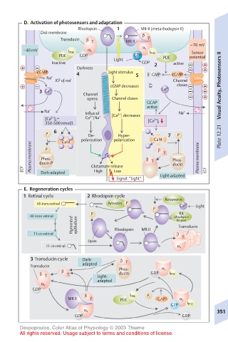
D.  Activation of photosensors and adaptation
                          Rhodopsin   1       MR-II (metarhodopsin II)
              Disk membrane
                                              β
                    Transducin  β  γ     MR II  γ
                                                             –70 mV
        –40 mV               α s              α s
                       I PDE                    I PDE         Sensor
                    PDE   GDP              GTP       PDE     potential
                                     Light
                 Inactive                     GDP  2    active
                         Darkness
             cGMP        4         Light stimulus  5‘-GMP  cGMP
                Na +                        5
                  ICF of rod                            Channel
                                    cGMP decreases  ICF  2+  closes    Visual Acuity, Photosensors II
             3  Ca 2+      Channel                  Ca
                             opens  Channel closes
                                               GCAP
                                               active
               Na +         Influx of  2+                Na +
                             2+
                 2+
               [Ca ] i  =   Ca /Na +  [Ca ] decreases  2+
               350-500 nmol/L                   [Ca ] i
                  6
               P             De-      Hyper-          7  P             Plate 12.21
                           polarization  polarization  CaM
                     CaM
           Plasma membrane  Phos-              β  γ  β  γ  Phos-  Plasma membrane
               P
        ECF  ducin-P          Glutamate release         ducin     ECF
                                     Low
                               High
              Dark-adapted
                                      Signal: “Light“  Light-adapted
         E.  Regeneration cycles
        1  Retinal cycle     2 Rhodopsin cycle
                                                     Recoverin
             All-trans-retinal     Arrestin  P            Ca 2+  Light
                                                 P
                              P                           RK
          All-trans-retinol                             (rhodopsin
                        Pigmented  epithelium  Rhodopsin  MR II  Transducin
                                                         kinase)
            11-cis-retinol                               β  γ
                             Opsin
                11-cis-retinal                           α s
           3 Transducin cycle  Dark-   β  γ
           Transducin      adapted
                    β                  Phos-        α s
              β  γ    γ         Light-  ducin    GDP   I PDE
                                adapted
              α s
           GDP
                            β             I PDE  P i
                       MR II  γ        PDE         GAP    α s
                                                       GTP  I PDE
                            α s                                       351
                         GDP                               GDP
       Despopoulos, Color Atlas of Physiology © 2003 Thieme
       All rights reserved. Usage subject to terms and conditions of license.