
Page 359 - Color_Atlas_of_Physiology_5th_Ed._-_A._Despopoulos_2003
P. 359
rior focal length in meters, and is measured in
Optical Apparatus of the Eye diopters (dpt). In accommodation for far vision,
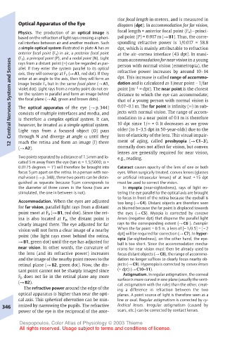
Physics. The production of an optical image is focal length = anterior focal point (F a)—princi-
based on the refraction of light rays crossing a spheri- pal point (P) = 0.017 m (! B1). Thus, the corre-
cal interface between air and another medium. Such sponding refractive power is 1/0.017 = 58.8
a simple optical system illustrated in plate A has an dpt, which is mainly attributable to refraction
anterior focal point (F a) in air, a posterior focal point at the air–cornea interface (43 dpt). In maxi-
(F p), a principal point (P), and a nodal point (N). Light
mum accommodation for near vision in a young
Central Nervous System and Senses enter at an angle to the axis, then they will form an dpt. This increase is called range of accommo-
rays from a distant point (!) can be regarded as par-
person with normal vision (emmetropia), the
allel. If they enter the system parallel to its optical
refractive power increases by around 10–14
axis, they will converge at F p (! A1, red dot). If they
dation and is calculated as 1/near point – 1/far
image beside F p but in the same focal plane (! A1,
– 1
point [m
violet dot). Light rays from a nearby point do not en-
= dpt). The near point is the closest
ter the system in parallel and form an image behind
distance to which the eye can accommodate;
the focal plane (! A2, green and brown dots).
that of a young person with normal vision is
0.07–0.1 m. The far point is infinity (!) in sub-
The optical apparatus of the eye (! p. 344)
jects with normal vision. The range of accom-
consists of multiple interfaces and media, and
modation to a near point of 0.1 m is therefore
is therefore a complex optical system. It can,
older (to 1–3.5 dpt in 50-year-olds) due to the
Light rays from a focused object (O) pass
loss of elasticity of the lens. This visual impair-
through N and diverge at angle α until they
ment of aging, called presbyopia (! C1–3),
reach the retina and form an image (I) there
12 however, be treated as a simple optical system. 10 dpt since 1/! = 0. It decreases as we grow
(! A2). normally does not affect far vision, but convex
lenses are generally required for near vision,
Two points separated by a distance of 1.5 mm and lo- e.g., reading.
cated 5 m away from the eye (tan α = 1.5/5000; α =
0.0175 degrees ! 1!) will therefore be brought into Cataract causes opacity of the lens of one or both
focus 5µm apart on the retina. In a person with nor- eyes. When surgically treated, convex lenses (glasses
mal vision (! p. 348), these two points can be distin- or artificial intraocular lenses) of at least + 15 dpt
guished as separate because 5 µm corresponds to must be used to correct the vision.
the diameter of three cones in the fovea (two are In myopia (near-sightedness), rays of light en-
stimulated, the one in between is not). tering the eye parallel to the optical axis are brought
to focus in front of the retina because the eyeball is
Accommodation. When the eyes are adjusted too long (! C4). Distant objects are therefore seen
for far vision, parallel light rays from a distant as blurred because the far point is displaced towards
point meet at F p (! B1, red dot). Since the ret- the eyes (! C5). Myopia is corrected by concave
ina is also located at F p, the distant point is lenses (negative dpt) that disperse the parallel light
clearly imaged there. The eye adjusted for far rays to the corresponding extent (! C6 ). Example:
vision will not form a clear image of a nearby When the far point = 0.5 m, a lens of [– 1/0.5] = [– 2
point (the light rays meet behind the retina, dpt] will be required for correction (! C7). In hyper-
opia (far-sightedness), on the other hand, the eye-
! B1, green dot) until the eye has adjusted for ball is too short. Since the accommodation mecha-
near vision. In other words, the curvature of nisms for near vision must then be already used to
the lens (and its refractive power) increases focus distant objects (! C8), the range of accommo-
and the image of the nearby point moves to the dation no longer suffices to clearly focus nearby ob-
retinal plane (! B2, green dot). Now, the dis- jects (! C9). Hyperopia is corrected by convex lenses
tant point cannot not be sharply imaged since (+ dpt) (! C10–11).
F p does not lie in the retinal plane any more Astigmatism. In regular astigmatism, the corneal
(! B2). surface is more curved in one plane (usually the verti-
cal: astigmatism with the rule) than the other, creat-
The refractive power around the edge of the ing a difference in refraction between the two
optical apparatus is higher than near the opti- planes. A point source of light is therefore seen as a
cal axis. This spherical aberration can be min- line or oval. Regular astigmatism is corrected by cy-
346 imized by narrowing the pupils. The refractive lindrical lenses. Irregular astigmatism (caused by
power of the eye is the reciprocal of the ante- scars, etc.) can be corrected by contact lenses.
Despopoulos, Color Atlas of Physiology © 2003 Thieme
All rights reserved. Usage subject to terms and conditions of license.