
Page 98 - Color_Atlas_of_Physiology_5th_Ed._-_A._Despopoulos_2003
P. 98
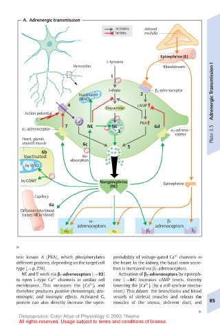
A.  Adrenergic transmission
                                       Activates  Adrenal
                                       Inhibits  medulla


                                                      Epinephrine (E)
                                    L-tyrosine                         I
                         Varicosities                 Bloodstream
                                     1                                 Transmission

                                     L-dopa         β 2 -adrenoceptor
                           Inactivated         2
                           (MAO)
                       4                      cAMP
                                    Dopamine                           Adrenergic
         Action potential
                          Ca 2+                PKA
                      7       NE      NE            6d
        α 2 -adrenoceptor               3                α 2 -adreno-
                                                          ceptor       Plate 3.5
        Heart, glands,
        smooth muscle       6c
                                           5
               6b
         Inactivated:       Re-
                        absorption
         by MAO
        by COMT                   Norepinephrine
                                     (NE)              Epinephrine
            Capillary
                 6a
        Diffusion into blood
        (raises NE in blood)
                              α-                        β-
                          adrenoceptors             adrenoceptors
                      α 1             α 2        β 1            β 2

      !
      tein kinase A (PKA), which phosphorylates  probability of voltage-gated Ca 2+  channels in
      different proteins, depending on the target cell  the heart. In the kidney, the basal renin secre-
      type (! p. 274).                tion is increased via ! 1-adrenoceptors.
        NE and E work via " 1-adrenoceptors (! B3)  Activation of " 2-adrenoceptors by epineph-
      to open L-type Ca 2+  channels in cardiac cell  rine (! B4) increases cAMP levels, thereby
                                                 2+
      membranes. This increases the [Ca ] i and  lowering the [Ca ] i (by a still unclear mecha-
                              2+
      therefore produces positive chronotropic, dro-  nism). This dilates the bronchioles and blood
                                                                  !
      motropic, and inotropic effects. Activated G s  vessels of skeletal muscles and relaxes the
      protein can also directly increase the open-  muscles of the uterus, deferent duct, and  85
                                                                  !
       Despopoulos, Color Atlas of Physiology © 2003 Thieme
       All rights reserved. Usage subject to terms and conditions of license.