
Page 496 - Hall et al (2015) Principles of Critical Care-McGraw-Hill
P. 496
366 PART 3: Cardiovascular Disorders
A
A
B
B C
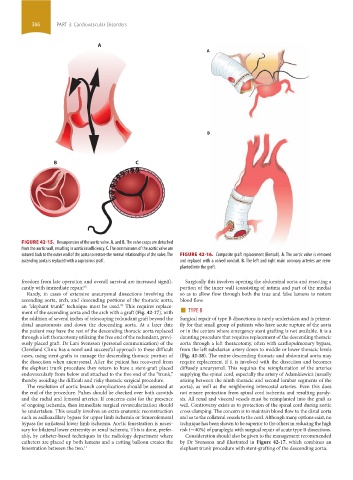
FIGURE 42-15. Resuspension of the aortic valve. A. and B. The valve cusps are detached
from the aortic wall, resulting in aortic insufficiency. C. The commissures of the aortic valve are
sutured back to the outer wall of the aorta to restore the normal relationships of the valve. The FIGURE 42-16. Composite graft replacement (Bentall). A. The aortic valve is removed
ascending aorta is replaced with a suprasinus graft. and replaced with a valved conduit. B. The left and right main coronary arteries are reim-
planted into the graft.
freedom from late operation and overall survival are increased signifi- Surgically this involves opening the abdominal aorta and resecting a
cantly with immediate repair. 65 portion of the inner wall (consisting of intima and part of the media)
Rarely, in cases of extensive aneurysmal dissections involving the so as to allow flow through both the true and false lumens to restore
ascending aorta, arch, and descending portions of the thoracic aorta, blood flow.
ment of the ascending aorta and the arch with a graft (Fig. 42-17), with ■ TYPE B
70
an “elephant trunk” technique must be used. This requires replace-
the addition of several inches of telescoping redundant graft beyond the Surgical repair of type B dissections is rarely undertaken and is primar-
distal anastomosis and down the descending aorta. At a later date ily for that small group of patients who have acute rupture of the aorta
the patient may have the rest of the descending thoracic aorta replaced or in the centers where emergency stent grafting is not available. It is a
through a left thoracotomy utilizing the free end of the redundant, previ- daunting procedure that requires replacement of the descending thoracic
ously placed graft. Dr Lars Svensson (personal communication) of the aorta through a left thoracotomy, often with cardiopulmonary bypass,
Cleveland Clinic has a novel and successful approach to these difficult from the left subclavian artery down to middle or lower thoracic levels
cases, using stent-grafts to manage the descending thoracic portion of (Fig. 42-18). The entire descending thoracic and abdominal aorta may
the dissection when aneurysmal. After the patient has recovered from require replacement if it is involved with the dissection and becomes
the elephant trunk procedure they return to have a stent-graft placed diffusely aneurysmal. This requires the reimplantation of the arteries
endovascularly from below and attached to the free end of the “trunk,” supplying the spinal cord, especially the artery of Adamkiewicz (usually
thereby avoiding the difficult and risky thoracic surgical procedure. arising between the ninth thoracic and second lumbar segments of the
The resolution of aortic branch complications should be assessed at aorta), as well as the neighboring intercostal arteries. Even this does
the end of the procedure. Pulses should be checked over both carotids not ensure protection from spinal cord ischemia and resulting paraly-
and the radial and femoral arteries. If concerns exist for the presence sis. All renal and visceral vessels must be reimplanted into the graft as
of ongoing ischemia, then immediate surgical revascularization should well. Controversy exists as to protection of the spinal cord during aortic
be undertaken. This usually involves an extra-anatomic reconstruction cross-clamping. The concern is to maintain blood flow to the distal aorta
such as axilloaxillary bypass for upper limb ischemia or femorofemoral and so to the collateral vessels to the cord. Although many options exist, no
bypass for unilateral lower limb ischemia. Aortic fenestration is neces- technique has been shown to be superior to the others in reducing the high
sary for bilateral lower extremity or renal ischemia. This is done, prefer- risk (∼40%) of paraplegia with surgical repair of acute type B dissections.
ably, by catheter-based techniques in the radiology department where Consideration should also be given to the management rec ommended
catheters are placed up both lumens and a cutting balloon creates the by Dr Svensson and illustrated in Figure 42-17, which combines an
fenestration between the two. 11 elephant trunk procedure with stent-grafting of the descending aorta.
section03.indd 366 1/23/2015 2:08:36 PM