
Page 492 - Hall et al (2015) Principles of Critical Care-McGraw-Hill
P. 492
362 PART 3: Cardiovascular Disorders
lumens, when the false lumen is thrombosed. Recent studies have also
demonstrated the added benefit of contrast administration during TEE
for accurate identification of IMHs, penetrating aortic ulcers, as well as
better assessment of flow dynamics within the dissected aorta. 48
TEE is generally well tolerated, can be performed under conscious
sedation at the bedside, or with minimal sedation or no sedation in
hemodynamically unstable patients. This procedure is, thus, preferable
49
to CT in the critically ill patient with suspected aortic dissection who
requires continuous ICU monitoring and is too unstable for transporta-
tion to the radiology suite.
MRI is very accurate, sensitive, and specific, but has very limited role
for the diagnosis of acute aortic dissection as the procedure requires
up to 45 minutes, which is difficult in a critically ill patient. 33,50,51 It is
not readily available on an emergent basis and is contraindicated in
patients with pacemakers, defibrillators, and various types of vascular
clips. Renal dysfunction is a relative contraindication when gado-
linium is used. For these reasons, it is mostly used in stable patients for
long-term follow-up, or when diagnosis is uncertain after CT or TEE.
Excellent contrast can be obtained between extraluminal structures,
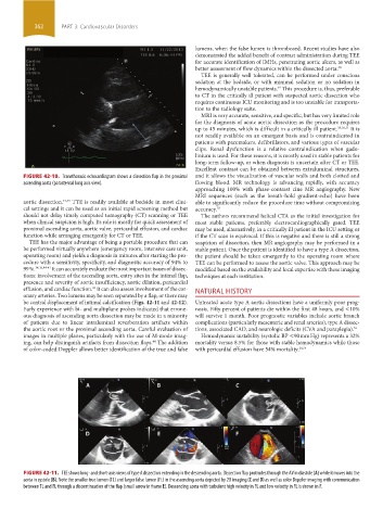
FIGURE 42-10. Transthoracic echocardiogram shows a dissection flap in the proximal and it allows the visualization of vascular walls and both clotted and
ascending aorta (parasternal long axis view). flowing blood. MR technology is advancing rapidly, with accuracy
approaching 100% with phase-contrast cine MR angiography. New
MRI sequences (such as the breath-hold gradient-echo) have been
aortic dissection. 42,43 TTE is readily available at bedside in most clini- able to significantly reduce the procedure time without compromising
cal settings and it can be used as an initial rapid screening method but accuracy. 52
should not delay timely computed tomography (CT) scanning or TEE The authors recommend helical CTA as the initial investigation for
when clinical suspicion is high. Its role is mostly for quick assessment of most stable patients, preferably electrocardiographically gated. TEE
proximal ascending aorta, aortic valve, pericardial effusion, and cardiac may be used, alternatively, in a critically ill patient in the ICU setting or
function while arranging emergently for CT or TEE. if the CT scan is equivocal. If this is negative and there is still a strong
TEE has the major advantage of being a portable procedure that can suspicion of dissection, then MR angiography may be performed in a
be performed virtually anywhere (emergency room, intensive care unit, stable patient. Once the patient is identified to have a type A dissection,
operating room) and yields a diagnosis in minutes after starting the pro- the patient should be taken emergently to the operating room where
cedure with a sensitivity, specificity, and diagnostic accuracy of 94% to TEE can be performed to assess the aortic valve. This approach may be
99 %. 34,36,44-47 It can accurately evaluate the most important issues of dissec- modified based on the availability and local expertise with these imaging
tions: involvement of the ascending aorta, entry sites in the intimal flap, techniques at each institution.
presence and severity of aortic insufficiency, aortic dilation, pericardial
effusion, and cardiac function. It can also assess involvement of the cor- NATURAL HISTORY
45
onary arteries. Two lumens may be seen separated by a flap, or there may
be central displacement of intimal calcification (Figs. 42-11 and 42-12). Untreated acute type A aortic dissections have a uniformly poor prog-
Early experience with bi- and multiplane probes indicated that errone- nosis. Fifty percent of patients die within the first 48 hours, and <10%
ous diagnosis of ascending aorta dissection may be made in a minority will survive 1 month. Poor prognostic variables include aortic branch
of patients due to linear intraluminal reverberation artifacts within complications (particularly mesenteric and renal arteries), type A dissec-
the aortic root or the proximal ascending aorta. Careful evaluation of tions, associated CAD, and neurologic deficits (CVA and paraplegia). 53
images in multiple planes, particularly with the use of M-mode imag- Hemodynamic instability (systolic BP <90 mm Hg) represents a 32%
ing, can help distinguish artifacts from dissection flaps. The addition mortality versus 8.5% for those with stable hemodynamics while those
44
of color-coded Doppler allows better identification of the true and false with pericardial effusion have 54% mortality. 28,54
FIGURE 42-11. TEE shows long- and short-axis views of type A dissection extending in the descending aorta. Dissection flap protrudes through the AV in diastole (A) while it moves into the
aorta in systole (B). Note the smaller true lumen (TL) and larger false lumen (FL) in the ascending aorta depicted by 2D imaging (C and D) as well as color Doppler imaging with communication
between TL and FL through a discontinuation of the flap (small arrow in frame E). Descending aorta with turbulent high velocity in FL and low velocity in FL is shown in F.
section03.indd 362 1/23/2015 2:08:28 PM