
Page 906 - Hall et al (2015) Principles of Critical Care-McGraw-Hill
P. 906
CHAPTER 69: Human Immunodeficiency Virus (HIV) and AIDS in the Intensive Care Unit 637
is also frequent. Bronchoscopic evaluation usually rules out a super- are meningitis, dementia, encephalopathy, focal neurologic deficits,
imposed treatable HIV-related disease in patients with pulmonary KS. myelopathy, peripheral neuropathy, and myopathy. 138,139 Often the neu-
Bronchoscopy and BAL may also allow visualization of the characteristic rologic disease may be associated with systemic illness rather than a
red-violaceous lesions in the endobronchial tree. Although biopsy of focal neurologic insult. The prevalence of CNS opportunistic infections
these bronchial lesions at times can provide diagnostic confirmation, is again dependent on the level of immune suppression. In addition to
this is rarely required and is not recommended due to concern regard- common viral and bacterial etiologies of meningoencephalitis, which
135
ing hemorrhage. Despite improvements in outcomes with ART-related can affect even immune-competent individuals, unusual infections
immune reconstitution and the use of chemotherapeutic regimens such such as Cryptococcus neoformans and Toxoplasma are AIDS-defining
as liposomal doxorubicin (alternatively paclitaxel), mortality of KS conditions. With advanced disease (CD4 cell counts <200 cells/mm )
3
remains high. 136,137 Corticosteroids may cause progression of cutaneous progressive multifocal leukoencephalopathy (PML) associated with JC
or visceral KS and are contraindicated. virus and primary CNS lymphomas must be considered although are
■ NEUROLOGIC MANIFESTATIONS IN HIV-INFECTED PATIENTS most commonly seen in those with CD4 cell counts <50 cells/mm .
3
The various etiologic agents responsible for these syndromes, in addi-
Neurologic disease secondary to opportunistic infection or neoplasm tion to key points of clinical presentation and diagnostic evaluation, are
in HIV-infected individuals may be associated with a depressed level summarized in Table 69-6. In general, most of the treatable infections
of consciousness and occasionally precipitates ICU care. The most complicating AIDS produce either meningitis or progressive focal neu-
frequently encountered neurologic syndromes in HIV-infected patients rologic deficits due to localized inflammatory lesions in the brain, often
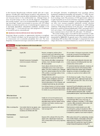
TABLE 69-6 Neurologic Complications in HIV-Infected Individuals
Neurologic Syndrome Etiologic Agents Clinical Presentation Diagnostic Evaluation
Meningitis
Aseptic (HIV) Headache, meningismus, and fever (all less CSF examination: mild mononuclear pleocytosis, elevated
common in chronic cases), with/without cranial protein, glucose normal (differential diagnosis also
neuropathies (V, VII, VIII); may occur with serocon- includes syphilis and lymphomatous meningitis)
version, but more common later in HIV disease
Bacterial (S pneumoniae, N meningiditis, Fever, headache with/without meningeal signs, CSF examination: polymorphonuclear pleocytosis, high
Listeria monocytogenes, H influenzae) confusion, seizures. Bacterial meningitis rare in protein, low glucose, with/without positive Gram stain
HIV-infected patients bacterial cultures of blood and CSF
M tuberculosis Usually subacute-chronic meningitis. Clinical CSF examination: lymphocytic pleocytosis, low glucose, and
manifestations similar to those in HIV-negative increased protein; smear for acid-fast-bacilli insensitive;
patients. Fever (89%), headache (59%), meningeal cultures for M tuberculosis required. Rapid CSF diagnosis
signs (65%), altered mentation (43%), focal deficits through the use of nucleic acid amplification assays is
(19%), and clinical or radiologic evidence of extra- promising, but assays have not been validated in CSF
meningeal tuberculosis (65%)
C neoformans Often headache, fever, and vomiting; sometimes CSF white blood count usually <20/µL; CSF, glucose,
confusion, seizure, meningismus, cranial nerve and protein often normal; cryptococcal antigen positive
palsies; occasionally meningitis symptoms are in CSF (>90%) and serum (94%-100%); 50% will have
minimal and presentation is fever, fungemia, and/ associated fungemia
or extrameningeal lesion (eg, skin, pneumonia)
Coccidioides immitis Fever, lethargy, headache, with/without meningis- Complement-fixing (CF) antibody titer positive in 83% of
mus, confusion (consider if travel/residence history patients with AIDS-related coccidioidomycosis. Any positive
for endemic zone, eg, southwestern United States). CSF titer of CF antibodies is usually diagnostic of meningitis.
CSF profile: lymphocytic pleocytosis usually >50 cells/µL,
elevated protein, low glucose. Fungal cultures of blood and CSF
Diffuse brain disease
HIV-associated dementia HIV Usually alert, but impaired cognition (usually Abnormalities on neurocognitive testing. Other findings
concentration and memory), behavior (apathy, may include hyperreflexia, ataxia, release signs, leg
personality change), and motor function (slowing weakness, incontinence, and mutism. CT or MRI: atrophy
and reduced coordination); sometimes organic ± patchy or diffuse abnormalities of hemispheric white
psychosis or mania matter seen on MRI (T2-weighted)
Diffuse encephalopathies Toxic metabolic disorders (eg, hypoxia, sepsis, Impaired alertness and cognition, with/without Blood chemistry to exclude metabolic causes, with/
drugs), CNS toxoplasmosis, CNS lymphoma, focal neurologic deficits without drug levels, serology for toxoplasmosis. MRI or
occasionally viral infection (CMV, HSV) contrast-enhanced CT head scan: focal lesions may be seen
in toxoplasmosis, lymphoma, herpes simplex encephalitis
Focal brain disease
Toxoplasmosis Toxoplasma gondii Headache (55%), confusion (52%), fever (47%), MRI or contrast-enhanced CT scan: spherical ring–enhancing
seizures (29%), reduced level of consciousness lesions in cortex, thalamus, or basal ganglia, but may have
(42%), and focal deficits (69%) usually progressing atypical appearances. Toxoplasma serum serology (IgG)
over days usually positive (84%); possible brain biopsy if no response
to empiric therapy for toxoplasmosis (see Fig. 69-1).
(Continued)
section05_c61-73.indd 637 1/23/2015 12:48:22 PM