
Page 938 - Hall et al (2015) Principles of Critical Care-McGraw-Hill
P. 938
CHAPTER 72: Encephalomyelitis 669
TABLE 72-2 Less Common Causes of Encephalitis, Diagnosis and Treatment Summary (Continued )
Etiology Epidemiology Clinical Features Diagnosis Treatment
Cysticercosis Mexico, Central and South Most common presentation is new onset Serology Decision to treat must be individualized
Taenia solium America, Southeast Asia seizures due to CNS cysticercosis CSF antibodies (negative test does not Albendazole and corticosteroids are
Acquired by ingestion of eggs Rarely encephalitic presentation with rule out the diagnosis) preferred
In humans larval stage causes CNS very high cyst burden in the brain or after CT/MRI of brain may show cystic lesions Praziquantel is an alternative
disease treatment with or without calcifications Surgical resection when indicated
Ring enhancement and edema may be seen
Baylisascaris Children often affected Unilateral neuroretinitis, meningoen- CSF and peripheral eosinophilia Albendazole plus diethylcarbamazine
procyonis Contact with dirt contaminated cephalitis Serology not readily available plus
with raccoon feces (playing in or High rates of permanent neurologic Larvae identification in tissue adjunctive corticosteroids should be
eating dirt) sequelae and mortality considered
MRI may show white matter lesions
Gnathostoma Southeast Asia and Latin America Eosinophilic meningoencephalitis (a less CSF—often xanthochromic or bloody Albendazole or ivermectin
spinigerum Ingestion of undercooked fish, common manifestation) with eosinophilic pleocytosis Addition of corticosteroids may be
frogs, eels, snakes, and poultry Sudden onset of headache, radicular Peripheral eosinophilia beneficial in suppressing inflammation
pain, parasthesias followed by paralysis, Worms identification in tissues
cranial nerve palsies, and bladder
Serology not readily available
Incontinence
survivors end up with significant neurologic deficits. Empiric ther- DNA can be detected in the CSF once therapy has been initiated
2
apy should be initiated even with minimal clinical suspicion before is unclear. Discontinuation of therapy based on negative CSF PCR
the onset of dominant temporal lobe hemorrhagic necrosis and sig- results depends on the clinical probability of HSVE and is a matter of
nificant deterioration of consciousness. The recommended treatment judgment. Use of corticosteroids is controversial though a few stud-
16
of HSVE is acyclovir at a dose of 10 mg/kg every 8 hours for 14 to ies have shown favorable outcomes. 17
21 days. It prevents viral replication by inhibiting the viral (as well In spite of effective treatment, mortality is still up to 20% to 30%. The
3
as the cellular) DNA polymerase in infected cells. Introduced in the precise factors that determine the therapeutic response are unknown.
mid-1980s, it replaced vidarabine and was shown to reduce mortality Patients with low initial level of consciousness and age over 30 have
from 70% to 20%. 13,14 Relapses occur commonly in children within 2 a very poor prognosis in general. Long-term complications of HSVE
weeks of completing antiviral therapy necessitating a second course infection are common and include neurocognitive impairment, residual
of acyclovir. Acyclovir has renal and neurologic toxicity manifesting dysphasias, paresis, paresthesias, behavioral changes, and a Korsakoff-
15
as crystalluria, renal failure, and encephalopathy. Effective hydration, like amnesia. 18
receiving acyclovir for suspected HSVE should also receive other ■ HERPES SIMPLEX VIRUS TYPE 2
dose adjustment, or discontinuation of therapy is helpful. Patients
broad-spectrum antibiotics for the first 48 to 72 hours until CSF and HSV-2 encephalitis is seen primarily in neonates, where brain involve-
other cultures for bacteria are negative. The time period that HSV ment is generalized and is usually acquired during vaginal delivery from
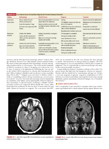
FIGURE 72-1. Axial FLAIR image of MRI of the brain showing increased signal intensity FIGURE 72-2. Coronal FLAIR of MRI of the brain showing increased signal intensity in
in bilaterial temporal lobes. bilateral temporal lobes.
section05_c61-73.indd 669 1/23/2015 12:48:44 PM