
Page 935 - Hall et al (2015) Principles of Critical Care-McGraw-Hill
P. 935
666 PART 5: Infectious Disorders
■
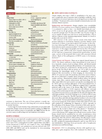
TABLE 72-1 Common Causes of Encephalitis HERPES SIMPLEX VIRUS ENCEPHALITIS
Viral Papovaviridae Herpes simplex virus type 1 (HSV-1) encephalitis is the most com-
Herpes viridae JC virus—progressive multifocal mon recognizable cause of sporadic fatal encephalitis worldwide. Early
Herpes simplex virus (HSV-1, HSV-2) leukoencephalopathy recognition is of utmost importance due to its significant mortality and
1,2
Varicella-zoster virus (VZV) Prion disease morbidity. Treatment with acyclovir should be initiated early even with
Cytomegalovirus (CMV) Creutzfeldt-Jakob disease (CJD) minimal clinical suspicion.
Epstein-Barr virus (EBV) Variant Creutzfeldt-Jakob disease Epidemiology and Pathogenesis: Herpes simplex virus encephalitis
Human herpes virus 6 (HHV-6) (vCJD) (HSVE) is the most common cause of fatal nonepidemic encephalitis.
Herpes B virus Bacteria It is estimated to affect at least 1 in 500,000 individuals per year.
1
Flaviviridae Bartonella species (henselae HSVE occurs in all age groups, with a bimodal peak distribution
West Nile virus and bacilliformis) in patients younger than 20 years and older than 50 years of age. It
Japanese encephalitis virus Listeria monocytogenes occurs equally in both sexes and has no racial predilection. This is
St Louis encephalitis virus Mycoplasma pneumoniae seldom a disease of the immunocompromised. Indeed, it is hard to
Powassan virus Mycobacteria predict who is at risk for HSVE. 3
Murray Valley encephalitis virus Mycobacterium tuberculosis HSV infection of the central nervous system arises from either
Tick-borne encephalitis virus Rickettsia and ehrlichioses primary infection or reactivation. Primary infection, which is most
Picornaviridae Rickettsia rickettsii (Rocky Mountain common in children, occurs due to direct CNS invasion via the olfac-
Polio virus spotted fever) tory tract following HSV infection of the oropharynx. Alternatively,
Nonpolio enteroviruses Anaplasma phagocytophilum (human latent infection most common in adults >50 years occurs due to viral
Echoviruses granulocytotrophic ehrlichiosis) reactivation in the trigeminal ganglia and reaches the CNS along a
Coxsackieviruses Ehrlichia chaffeensis (human neurotropic route. HSVE is the most common clinical presentation
Numbered enteroviruses monocytotrophic ehrlichiosis) of HSV and has a characteristic temporal lobe predilection. In most
Bunyaviridae Coxiella burnetii (Q fever) cases, necrosis of the temporal lobes is seen with associated clinical
California encephalitis group Spirochetes deficits. Much of the pathogenesis of HSVE seems to be immune
La Crosse virus Borrelia burgdorferi (Lyme disease) mediated. 4,5
Jamestown Canyon virus Treponema pallidum (syphilis)
Togaviridae Fungi Clinical Features and Diagnosis: There are no typical clinical features of
Eastern equine encephalitis virus Coccidioides species HSVE. The clinical syndrome is often characterized by acute onset of
Western equine encephalitis virus Cryptococcus neoformans fever, headache, seizures, focal neurologic deficits, and altered mental
Venezuelan equine encephalitis virus Histoplasma capsulatum status. There may be an influenza-like prodrome. CSF examination typi-
Rubella virus Protozoa cally shows lymphocytic predominance, elevated protein, and normal
Rhabdoviridae Toxoplasma gondii glucose levels. Red blood cells are present in 75% to 80% of samples,
6
Rabies virus Acanthamoeba species indicating the hemorrhagic nature of this encephalitis. Unilateral
Orthomyxoviridae Balamuthiamandrillaris temporal lobe abnormalities on brain imaging are characteristic for
3
Influenza virus Naegleria fowleri HSVE. CT scans have only 50% sensitivity early in the course of illness,
Paramyxoviridae Plasmodium falciparum (malaria) but can be performed to exclude raised intracranial pressure prior to
Measles virus Trypanosoma bruceigambiense (West lumbar puncture. Abnormalities include localized edema, low-density
Mumps virus African trypanosomiasis) lesions, mass effect, contrast enhancement, and hemorrhage. On the
Nipah virus Trypanosoma bruceirhodesiense (East other hand, MRI is the most sensitive and specific neurodiagnostic test
Hendra virus African trypanosomiasis) for HSVE and has excellent delineation of the temporobasal lobe of
Retroviridae Helminths the brain (Figs. 72-1 and 72-2). Diffusion-weighted MRI is especially
7,8
Human immune deficiency virus Baylisascaris procyonis preferred early in the course of the disease (Fig. 72-3). MRI shows
Human T-cell lymphotropic virus Gnathostoma species abnormal signal earliest in FLAIR sequences and diffusion-restricted
Adenoviridae Taenia solium (cysticercosis) images of the medial temporal lobes and insulas. Brain perfusion SPECT
Adenovirus Postinfectious scan shows increased tracer accumulation in the affected temporal lobe
9
Poxviridae Acute disseminated encephalomyelitis with high specificity. Focal EEG findings occur in 80% to 90% of cases
Vaccinia virus of HSVE, but are nonspecific. The EEG typically shows focal spiked and
slow wave (theta and delta slowing) patterns localized to the area of the
brain involved. Paroxysmal lateralized epileptiform discharges are also
continues to deteriorate. The care of these patients is mostly sup- seen but are nonspecific. PCR analysis to detect HSV DNA in CSF is the
portive and recovery can occur after long and even extreme changes gold standard for establishing the diagnosis of HSVE, especially in
in mental function. the early phase of the disease. It has a sensitivity of 98% and a specific-
A wide range of viruses, bacteria, fungi, and parasites are associated with ity of 99%. In very early cases, PCR can be negative and it also becomes
encephalitis, though viruses are the most common etiology. See Table 72-1 negative by the second or third week of the disease. 10,11 Treatment of
for the most common causes of encephalitis, and Table 72-2 for the less HSVE should be initiated while awaiting PCR results. Later in the
common causes, as well as diagnostic and treatment information. course of the disease, HSV antigen and antibody become positive. Brain
biopsy, should only be considered in very select circumstances, when
HERPES VIRIDAE the diagnosis remains uncertain despite all available investigations and
particularly when the patient is deteriorating clinically despite empiric
Viruses that are associated with encephalomyelitis in the Herpesviridae therapy. Biopsy specimens are tested for presence of HSV by culture,
12
family are herpes simplex virus 1 and 2 (HSV-1 and HSV-2), varicella- for HSV antigens by immunohistochemistry, and for viral DNA by in
zoster virus (VZV), cytomegalovirus (CMV), Epstein-Barr virus (EBV), situ hybridization. Pathology shows perivascular cuffs of lymphocytes
human herpesvirus 6 (HHV-6), and herpes B virus. Viral shedding and numerous small hemorrhages.
occurs in symptomatic and asymptomatic persons infected with HSV,
CMV, EBV, and HHV-6 and can be transmitted by virus-containing Management: HSVE is a devastating infection, with significant mor-
body fluids to susceptible hosts. tality and a grim prognosis. Even with treatment two-thirds of the
section05_c61-73.indd 666 1/23/2015 12:48:43 PM