
Page 946 - Hall et al (2015) Principles of Critical Care-McGraw-Hill
P. 946
CHAPTER 73: Life-Threatening Infections of the Head, Neck, and Upper Respiratory Tract 677
physicians unfamiliar with these entities may underestimate their extent
• The development of marked asymmetry in the course of a and severity. In this chapter, the key clinical manifestations of several
submandibular space infection should be viewed with great con- life-threatening infections of the head, neck, and upper respiratory tract
cern, since it may be indicative of extension to the lateral pharyn- are highlighted, and the critically important anatomic relationships that
geal space. underlie their diagnosis and management are emphasized.
• In immunocompromised patients, the classical manifestations of
infection, such as edema and fluctuance at the local site and fea- GENERAL ANATOMIC CONSIDERATIONS
tures of systemic toxicity, may be absent. Life-threatening infections of the head, neck, and upper respiratory tract
• β-lactam-β-lactamase inhibitor or penicillin in combination with most commonly originate from suppurative complications of dental,
metronidazole is the antibiotic regimen of choice for odontogenic oropharyngeal, or otorhinolaryngeal infections. From these sites, infec-
deep neck infections, but immunocompromised patients require a tion may extend along natural fascial planes into deep cervical spaces
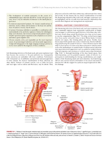
broader-spectrum against organisms such as Staphylococcus aureus or vascular compartments (Fig. 73-1). The deep cervical fascia ranges
1
and enteric gram-negative rods. from loose areolar connective tissue to dense fibrous bands. It invests
• Chronic sinusitis, otitis, and mastoiditis are the most important muscles and organs, thus forming planes and spaces. Notably, these
causes of parameningeal infection and intracranial suppuration. fascial planes both separate and connect distant areas, thereby both lim-
Computed tomography is the single imaging technique proven to iting and directing the spread of infection. These infections may be fatal
be the most useful for the diagnosis of these conditions. either by local airway occlusion or by direct extension to vital structures
such as the mediastinum or carotid sheath. Otorhinocerebral infections
may cause intracranial suppuration such as cerebral or epidural abscess,
subdural empyema, and cavernous or cortical venous sinus thrombosis
Life-threatening infections of the head, neck, and upper respiratory tract (Fig. 73-2). A thorough knowledge of the deep fascial spaces, their
2
have become less common in the post-antibiotic era. Consequently, interrelationships, and the potential anatomic routes of infection is a
many physicians are unfamiliar with these conditions. Furthermore, prerequisite to understanding the etiology, manifestations, and compli-
with widespread use of antibiotics and profound immunosuppression cations of life-threatening head and neck infections. Such knowledge
in some patients, the classical manifestations of these infections are will not only provide valuable information on the nature and extent of
often altered. Features of systemic toxicity, such as chills and fever, infection but will also suggest the optimum surgical approach for effec-
and local signs, such as edema and fluctuance, may be absent. Thus, tive drainage.
A B
Pharyngeal
wall Parotid
gland
Tonsil Lateral pharyngeal
space
Hyoid
3 Hyoid Internal carotid artery
4 Sternohyoid m. Internal jugular vein
5
Sternothyroid
C6 Thyroid gland C Anterior
2 longitudinal
Sternum ligament 1
Anterior
longitudinal
ligament
3
4
1 1
5 C6 Esophagus
Layers of deep cervical fascia
Superficial Carotid sheath
Deep
Middle Platysma 1
membrane Trachea
Sternothyroid Thyroid
membrane 2 Sternohyoid
membrane gland
FIGURE 73-1. Relation of lateral pharyngeal, retropharyngeal, and prevertebral spaces to the posterior and anterior layers of deep cervical fascia: 1, superficial space; 2, pretracheal space;
3, retropharyngeal space; 4, “danger” space; 5, prevertebral space. A. Midsagittal section of the head and neck. B. Coronal section in the suprahyoid region of the neck. C. Cross section of the neck
at the level of thyroid isthmus. (Reproduced with permission from Chow AW. Infections of the oral cavity, neck and head. In: Mandell GL, Bennett JE, Dolin R. Principles and Practice of Infectious
Diseases. 7th ed. Philapelphia, PA: Elsevier Churchill Livingstone, Inc; 2010:855-871.)
section05_c61-73.indd 677 1/23/2015 12:48:52 PM