
Page 951 - Hall et al (2015) Principles of Critical Care-McGraw-Hill
P. 951
682 PART 5: Infectious Disorders
drainage is the most important mechanism of spread, it most often
involves the carotid sheath alone. A history of sore throat, while usually
present on admission, is not invariable; it may only be mild or unilateral,
and there may be a latent period of up to 3 weeks before manifestations
of deep infection develop. The patient presents either in a toxic condition
or insidiously with a fever of undetermined origin. Trismus is absent,
and signs of local suppuration may be subtle clinically because of the
tight connective tissue around and within the carotid sheath. This bar-
rier confines the infection and may limit it to only the internal jugular
vein. Dyspnea may be prominent as edema and swelling descend directly
to involve the epiglottis and larynx. Swelling of the pharyngeal wall, if
present, will be behind the palatopharyngeal arch and is easily missed.
Suppurative jugular thrombophlebitis (Lemierre syndrome) is the most
common vascular complication of a lateral pharyngeal space infection. 11,12
An indurated swelling a few centimeters long may be palpable behind the
sternocleidomastoid muscle or may be found more deeply behind the
palatopharyngeal arch. Trismus is minimal and may be absent. Vocal
cord paralysis or other neurologic signs representing lower cranial nerve
involvement may be present. These signs are frequently missed unless
specifically sought and may be transient. The patient may thus present
with sepsis but no obvious source (50% of cases). Metastatic abscesses
are common, characteristically involving the lungs, bones, and joints or
other sites. There may be retrograde spread of infection with cerebral
abscess or meningitis. A diagnosis of right-sided bacterial endocarditis
may be considered. In common with other anaerobic septic conditions,
hepatic enlargement, tenderness, abnormal liver function tests, and even
frank jaundice may be present, which may misdirect investigations and
further delay diagnosis. Positive gallium or white-cell–labeled indium
13
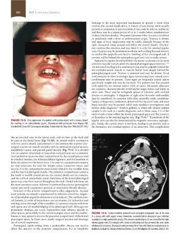
uptake in the neck is a useful diagnostic aid in these cases. CT of the
neck reveals edema within the lateral pharyngeal space and the presence
of thrombus in the internal jugular vein (Fig. 73-9). Thrombosis of the
37
FIGURE 73-8. Early appearance of a patient with Ludwig angina with a brawny, board- jugular vein can also be demonstrated by magnetic resonance angiogra-
like swelling in the submandibular spaces. (Reproduced with permission from Megran DW, phy. Rarely, the carotid artery is involved, leading to an arteritis and to
Scheifele DW, Chow AW. Odontogenic infections. Pediatr Infect Dis. May-June 1984;3(3):257-265.) the formation and eventual rupture of an aneurysm. This complication
like an inverted cone in the lateral neck, with its base at the skull and
its apex at the hyoid bone (Fig. 73-1B). Its medial wall is continuous
with the carotid sheath, and anteriorly it lies between the superior pha-
ryngeal constrictor muscle medially and the internal pterygoid muscle,
mandibular ramus, and parotid gland laterally (Fig. 73-6). It is divided
into an anterior (prestyloid or muscular) compartment and a posterior
(retrostyloid or neurovascular) compartment by the styloid process and
its attached muscles, the stylomandibular ligament, and the insertion of
these structures into the hyoid bone. The anterior compartment contains
no vital structures, but only fat, lymph nodes, connective tissue, and
muscle. It is the compartment most closely related to the tonsillar fossa
and the internal pterygoid muscle. The posterior compartment contains
the ninth to twelfth cranial nerves, the carotid sheath and its contents,
and the cervical sympathetic trunk. Infections of the lateral pharyngeal
space may arise from sources throughout the neck. Dental infections are
the most common source, followed by peritonsillar abscess (postanginal
sepsis) and rarely suppurative parotitis or mastoiditis (Bezold abscess).
Infection of the anterior compartment is often suppurative. Because
most patients are already compromised by infection elsewhere, diagno-
sis of lateral pharyngeal involvement is often delayed. The cardinal clini-
cal features, in order of importance, are (a) trismus, (b) induration and
swelling below the angle of the mandible, (c) systemic toxicity with fever
and rigors, and (d) medial bulging of the pharyngeal wall. Although not
prominent, dyspnea can occur. Suppuration may advance quickly to
other spaces, particularly to the retropharyngeal space and the medias- FIGURE 73-9. Contrast-medium enhanced axial computed tomographic scan of the neck
tinum, or may spread to involve the posterior compartment of the lateral in a young adult with jugular venous thrombosis–associated lateral pharyngeal space infection
pharyngeal space. In these cases, timely surgical incision and drainage secondary to a right peritonsillar abscess. The common carotid arteries (C) are normal but the right
are of utmost importance. internal jugular vein (J) is enlarged with a dense or enhancing wall that surrounds the more lucent
Postanginal sepsis arising from a peritonsillar abscess can involve intraluminal clot (arrow). (Reproduced with permission from Chow AW. Head and neck infections. In:
either the anterior or the posterior compartment, but as lymphatic Baddour L, Gorbach SL. Therapy of Infectious Diseases. 1st ed. Philadelphia, PA: Saunders; 2003:25-39.)
section05_c61-73.indd 682 1/23/2015 12:49:09 PM