
Page 948 - Hall et al (2015) Principles of Critical Care-McGraw-Hill
P. 948
CHAPTER 73: Life-Threatening Infections of the Head, Neck, and Upper Respiratory Tract 679
A space, the lateral pharyngeal space, and the retropharyngeal, “danger,”
and prevertebral spaces (Figs. 73-1, 73-5, and 73-6). Their salient
36
clinical features are summarized in Table 73-1. The potential pathways
of extension of these infections from one space to another are illus-
trated in Figure 73-7. The approach to radiographic and microbiologic
6
a b diagnosis is discussed toward the end of this chapter. Recommended
g
antimicrobial regimens for initial empirical therapy are summarized in
Table 73-2.
c ■ SUBMANDIBULAR SPACE INFECTIONS
f The prototypical infection of this space is known as Ludwig angina. In
1836, von Ludwig described five patients with “gangrenous induration
of the connective tissues of the neck, which advances to involve the
tissues that cover the small muscles between the larynx and the floor
of the mouth.” The infection is characteristically an aggressive, rapidly
d
spreading “woody” or brawny cellulitis involving the submandibular
g space. Although the submandibular space is divided by the mylohyoid
muscle into the sublingual space above and the submylohyoid space
e
below (Fig. 73-5), it can be considered a single unit owing to a direct
communication around the posterior aspect of the mylohyoid muscle.
Thus, classical Ludwig angina is a bilateral infection involving both
B the submylohyoid as well as the sublingual spaces. Ludwig angina
most commonly follows infection of the second or third mandibular
molar teeth (70%-85% of cases). The submylohyoid space is initially
involved, as the roots of these teeth are located below the attachments
of the mylohyoid muscle to the mandible (Fig. 73-4). Also, since the
lingual aspects of periodontal bone around these teeth are thinner,
a
medial spread of infection is facilitated. Infection extends contiguously
b (rather than by the lymphatics which would limit the infection to one
side) to involve the sublingual and thus the entire submandibular space
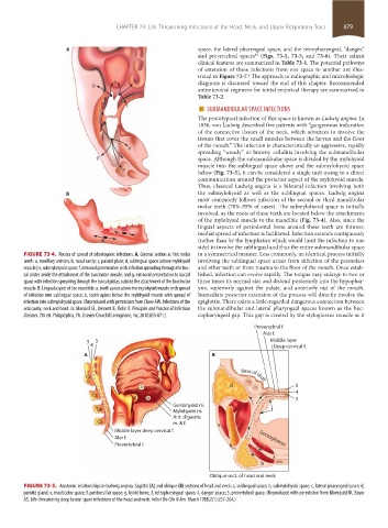
FIGURE 73-4. Routes of spread of odontogenic infections. A. Coronal section at first molar in a symmetrical manner. Less commonly, an identical process initially
teeth: a, maxillary antrum; b, nasal cavity; c, palatal plate; d, sublingual space (above mylohyoid involving the sublingual space arises from infection of the premolars
muscle); e, submylohyoid space; f, intraoral presentation with infection spreading through the buc- and other teeth or from trauma to the floor of the mouth. Once estab-
cal plates inside the attachment of the buccinator muscle; and g, extraoral presentation to buccal lished, infection can evolve rapidly. The tongue may enlarge to two or
space with infection spreading through the buccal plates outside the attachment of the buccinator three times its normal size and distend posteriorly into the hypophar-
muscle. B. Lingual aspect of the mandible: a, tooth apices above the mylohyoid muscle with spread ynx, superiorly against the palate, and anteriorly out of the mouth.
of infection into sublingual space; b, tooth apices below the mylohyoid muscle with spread of Immediate posterior extension of the process will directly involve the
infection into submylohyoid space. (Reproduced with permission from Chow AW. Infections of the epiglottis. There exists a little-regarded dangerous connection between
oral cavity, neck and head. In: Mandell GL, Bennett JE, Dolin R. Principles and Practice of Infectious the submandibular and lateral pharyngeal spaces known as the buc-
Diseases. 7th ed. Philapelphia, PA: Elsevier Churchill Livingstone, Inc; 2010:855-871.) copharyngeal gap. This gap is created by the styloglossus muscle as it
Prevertebral f.
Alar f.
5 3 Middle layer
4 Deep cervical f.
A B
C2 Base of skull
a d 5
4
C3 g b 3
Geniohyoid m.
C4 Mylohyoid m. f
Ant. digastric
C5 m. & f. e
Middle layer deep cervical f.
Alar f. c Genioglossus
Prevertebral f.
a
b
Oblique sect. of head and neck
FIGURE 73-5. Anatomic relationships in Ludwig angina. Sagittal (A) and oblique (B) sections of head and neck: a, sublingual space; b, submylohyoid space; c, lateral pharyngeal space; d,
parotid gland; e, masticator space; f, peritonsillar space; g, hyoid bone; 3, retropharyngeal space; 4, danger space; 5, prevertebral space. (Reproduced with permission from Blomquist IK, Bayer
AS. Life-threatening deep fascial space infections of the head and neck. Infect Dis Clin N Am. March 1988;2(1):237-264.)
section05_c61-73.indd 679 1/23/2015 12:49:04 PM