
Page 962 - Hall et al (2015) Principles of Critical Care-McGraw-Hill
P. 962
CHAPTER 74: Soft Tissue Infections 693
FIGURE 74-6. Necrotizing fasciitis with unopposed passage of a blunt instrument along the fas-
cial cleft, indicating the characteristic undermining between the subcutaneous tissue and deep fascia.
a sterile instrument along the plane just superficial to the deep fascia
(Fig. 74-6); the instrument cannot be passed with ordinary cellulitis.
Management: Before antimicrobial therapy is started, samples for
immediate Gram stain and for aerobic and anaerobic cultures should
be obtained by direct needle aspiration of the involved area. Probing
the lesion through an existing drainage site or through a small skin
incision will reveal the characteristic undermining of skin seen in
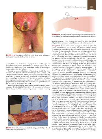
FIGURE 74-4. Fournier gangrene. Patches of necrotic skin are present on the scrotum necrotizing fasciitis. The use of full-thickness skin biopsy with frozen
35,36
and dusky erythema is present in the perineum and scrotum. section may aid the diagnosis, although some authors suggest that
this approach may not be practical in many settings. 1,37 Imaging stud-
ies using computed tomography and magnetic resonance imaging can
be very valuable to assess the presence and extent of the soft tissue
quickly followed by frank cutaneous gangrene. If an exudate is present, involvement, 23,24 but the use of imaging studies may be limited in
it may be serosanguineous and foul smelling. Systemic toxicity with dis- severely septic patients due to motion artifact and the valuable time
orientation is often severe. Large extracellular fluid shifts, hypotension, it takes in transporting an unstable patient for imaging studies 1,31,37 A
shock, and jaundice may follow. scoring system termed the Laboratory Risk Indicator for Necrotizing
38
Recently, a more indolent form of necrotizing fasciitis has been Fasciitis (LRINEC) was suggested as a diagnostic tool for necrotiz-
described, and the term subacute necrotizing fasciitis has been used. ing fasciitis, based on the values of multiple laboratory parameters for
The process is much slower and the clinical presentation evolves slowly patients presenting with soft tissue infections but was based on a retro-
over weeks to months with a slowly progressing soft tissue infection spective cohort and has not been substantiated in validation studies. 37
with minimal pain and discomfort. A variant of necrotizing fasciitis, The principles of management include general supportive measures,
31
involving the perineum, scrotum or penis, or vulva is known as Fournier administration of antimicrobial agents, and definitive surgery. General
gangrene and has a similar presentation but the findings are focused on measures include the placement of central venous and arterial monitor-
the perineal and genital areas (Fig. 74-4). 34 ing catheters, administration of intravenous fluids to correct dehydra-
A significant manifestation of necrotizing fasciitis is extensive under- tion, maintenance of adequate oxygenation, treatment of any underlying
mining of the skin (Fig. 74-5) associated with necrosis of subcutaneous diseases (eg, correction of ketoacidosis or congestive heart failure), and
fat and deep fascia. 1,5,31 The undermining can be demonstrated by passing attention to the patient’s nutritional needs. Enteral, and occasionally
parenteral, nutrition is required in the postoperative state to meet the
dramatically increased nitrogen requirements associated with tissue repair,
hyperthermia, sepsis, and vital organ requirements. Antibiotic selection
may be guided by the initial Gram stain, if available. Unfortunately there
are few, if any, randomized controlled trials to guide antimicrobial selec-
tion in necrotizing fasciitis and in the absence of specific microbiologic
data, broad-spectrum coverage should be given promptly, including
coverage for anaerobes, especially B. fragilis. An aminoglycoside (gen-
tamicin or tobramycin), 3 to 5 mg/kg per day in three divided doses
or, alternatively, 5 to 7 mg/kg as a single daily dose, plus clindamycin,
1200 to 2400 mg/day in three or four divided doses, is adequate initial
therapy for patients in whom renal function is not compromised. If large
gram-positive rods are noted on smear, suggesting clostridia, or if for
some reason clostridial infection is clinically suspected or if group A
Streptococcus is suspected, penicillin G should be added (20-24 million
U/day in divided doses). The combination of clindamycin and penicillin
is considered the treatment of choice for severe soft tissue infection due
to group A Streptococcus. In patients whose renal function is impaired
or rapidly changing owing to underlying disease or acute tubular
necrosis, a third-generation cephalosporin, such as cefotaxime, ceftri-
axone, or ceftazidime, or a fluoroquinolone can be used in place of the
FIGURE 74-5. Postoperative appearance of the lower leg presented in Figure 74–3. All aminoglycoside for gram-negative coverage. Alternatively, a carbapenem
necrotic subcutaneous tissue was excised. such as imipenem, meropenem, or doripenem may be used as a single
section05_c74-81.indd 693 1/23/2015 12:37:25 PM