
Page 963 - Hall et al (2015) Principles of Critical Care-McGraw-Hill
P. 963
694 PART 5: Infectious Disorders
agent. 1,7,18,37 In settings where community associated MRSA is suspected, fistula. Predisposing factors include diabetes mellitus, obesity, advanced
vancomycin (2 g/day in two divided doses) or another agent with reli- age, renal disease, and local trauma; diabetes mellitus is reported most
able activity against MRSA in skin and soft tissue infections, including commonly. With myonecrosis due to group A streptococci, no predis-
linezolid, daptomycin, or ceftaroline is indicated. In penicillin-allergic posing factors may be present. In drug addicts, infections of the extremi-
7,18
patients, metronidazole or chloramphenicol is useful as an alternative ties are more common, whereas perineal and buttock infections are
anaerobic agent. The addition of intravenous immunoglobulins, 0.4 g/kg more common in other populations.
per day for 4 to 5 days or 2 g/kg as a single dose with a repeat dose in When multiple microorganisms are responsible for myonecrosis, the
48 hours if the patient remains unstable, may be a useful adjunct for facultative bacteria assist the growth of anaerobes by using available
streptococcal toxic shock syndrome. 7,37,39-41 oxygen and destroying tissue (reducing the redox potential), which
The mainstay of management is surgical exploration, debridement, promotes a favorable milieu for the proliferation of anaerobic organ-
and drainage, which should be done as soon as possible. 1,7,37 Debridement isms. The process often involves muscle and fascia extensively, and it
and excision of all necrotic subcutaneous adipose tissue and fascia is may secondarily involve areas of subcutaneous tissue and skin. It should
required. The wound should be packed open. Daily exploration under be noted that necrotizing fasciitis will ultimately involve muscle, if left
general anesthesia is indicated for truncal or perirectal infections and for to progress.
all patients who remain in a toxic condition. Frequent dressing changes
are performed after suitable analgesia and are continued until healthy Etiology: Clostridium perfringens is the most common cause of clos-
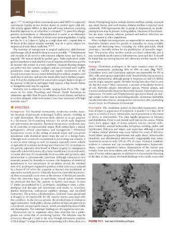
granulation tissue appears. Careful and regular reinspection of the tridial myonecrosis, producing 80% to 95% of cases (Fig. 74-7).
wound is necessary because initial debridement is seldom complete, and Clostridium novyi and Clostridium septicum are responsible for 5% to
small foci of infection and necrotic tissue often lead to further progres- 20%, with other species implicated rarely. Nonclostridial myonecrosis is
sion. It must be emphasized that conservative surgery leads to relapse of usually polymicrobial, although group A Streptococcus and CA-MRSA
the process. In the pelvic and upper thigh regions, a hip disarticulation may be single causative agents, the latter having been described in only
44
or hemipelvectomy may be required. the last few years. Most commonly, a mixture of facultative bacteria
Mortality rate is extremely variable, ranging from 4% to 74%. High (E coli, Klebsiella species, Enterobacter species, Proteus species, and
scores on the Acute Physiology and Chronic Health Evaluation on S aureus) and anaerobic bacteria (Bacteroides species, Peptostreptococcus,
admission, age older than 50 years, diabetes, truncal disease, and failure Peptococcus, Prevotella and Porphyromonas species) is found, an etiol-
to achieve adequate initial debridement have been associated with high ogy similar to that seen in necrotizing fasciitis. Aeromonas hydrophila
mortality rates. 42 has also been described as causing severe myonecrosis after penetrating
muscle injury in a freshwater environment.
■ MYONECROSIS Presentation: The incubation period of clostridial myonecrosis, from
Pathogenesis: The bacterial myonecrotic syndromes involve bacte- time of injury to appearance of symptoms, is usually 2 to 3 days, but it
rial invasion of previously undamaged healthy muscle, resulting in may be as brief as 6 hours. Intense pain, out of proportion to the extent
its rapid destruction. The process often referred to as gas gangrene of injury, is characteristic. The pain rapidly progresses in intensity
is a fulminant, life-threatening infection for which early diagnosis and distribution. Fever is not present until later in the course. Within
and intervention are essential. Bacterial myonecrotic syndromes may hours there appear signs of severe systemic toxicity: mental confu-
be of clostridial or nonclostridial origin. Both entities have a similar sion, irritability, marked tachycardia, tachypnea, sweating, pallor, and
pathogenesis, clinical presentation, and management. Clostridial hypotension. Delirium and stupor may supervene, although a period
5,7
myonecrosis occurs in the setting of muscle injury and concurrent of intense mental alertness may occur before the onset of delirium.
inoculation with clostridial spores from the soil or a foreign body. Renal failure, progressive hypotension and septic shock, intravascular
Although most commonly encountered in penetrating war wounds, it hemolysis, and disseminated intravascular coagulopathy may ensue.
is seen in other settings as well: (a) trauma, especially motor vehicle Bacteremia occurs in only 10% to 15% of cases. Profound metabolic
or agricultural accidents involving open fractures; (b) the postopera- acidosis is common and can overwhelm compensatory hyperventi-
tive period, especially after bowel or biliary surgery; (c) malignancy, lation, causing respiratory failure. Examination of the wound may
especially colorectal tumors; (d) arterial insufficiency in an extremity; initially show only tense edema and mild erythema. Later a spreading
(e) septic abortion; (f) occasionally, burn wounds; and (g) rarely, after zone of woody edema appears, in addition to a characteristic bronzing
intravascular or intramuscular injections. Although colonization of a of the skin. A thin, watery, brownish discharge with a sickly sweet odor
traumatic wound by clostridia is common, the frequency of clostridial
myonecrosis is very uncommon. In an animal model, the minimal
dose of C. perfringens required to produce a fatal infection is reduced
by a factor of 10 when the organism is injected into devitalized, as
6
opposed to normal, muscle. Clinically, however, clostridial myonecro-
sis does occasionally occur even in the absence of devitalized muscle.
Once the clostridia begin to proliferate, several potent exotoxins
are produced that have the capacity to destroy host tissue. At least
17 toxins are produced by C. perfringens, including α toxin, a phos-
pholipase that disrupts cell membranes and results in hemolysis,
platelet destruction, widespread capillary damage, and myofibril
destruction. The μ toxin, a hyaluronidase, facilitates tissue spread and
is thought to be responsible for the massive edema associated with
this condition. As the process spreads, the involved muscle undergoes
rapid destruction. Early pallor, edema, and loss of elasticity give way to
a discolored, noncontractile muscle, which eventually becomes friable
and disintegrates. The histologic findings are of coagulation necrosis.
Myonecrosis due to organisms other than clostridia has a patho-
genesis not unlike that of necrotizing fasciitis. The infection may be
introduced through a break in the skin, through intravenous injection
of illicit drugs, a surgical wound or enterostomy, a decubitus ulcer, or a FIGURE 74-7. Gram stain of Clostridium perfringens.
43
section05_c74-81.indd 694 1/23/2015 12:37:25 PM