
Page 958 - Hall et al (2015) Principles of Critical Care-McGraw-Hill
P. 958
CHAPTER 74: Soft Tissue Infections 689
of a classification of these entities is required, but, unfortunately, the Normally, the skin has a resident and a transient flora. The resident
published literature in this area may be confusing because of a lack of flora includes both bacteria and fungi but bacteria are most prevalent.
uniformity in descriptive terminology and the use of different classifi- The gram-positive cocci, including corynebacteria, propionibacteria,
cation schemes. The confusion is compounded by the fact that certain coagulase-negative staphylococci, Micrococcus, streptococci, lactococci,
clinical entities may involve one or more anatomic planes within the and Bacillus make up over 75% of the skin flora. Certain areas of the
subcutaneous tissue, and one or more bacterial species may be respon- body such as the buttocks, perineum, fossae and web spaces between
sible for the same or different clinical entities. Although classification the digits contain a more diverse flora and some gram-negative bacte-
schemes based on microbial etiology may be the most complete, they ria may be found including Acinetobacter, Serratia, Pseudomonas, and
offer little to the clinical diagnostic process necessary to expedite appro- occasionally anaerobic gram-negative bacteria. Staphylococcus aureus
3,4
priate management. To place a useful clinicoanatomic classification into is not considered part of the resident flora, but colonization rates of
1
perspective, a review of the basic anatomy and microbial ecology of the 10% to 30% in the anterior nares, axillae, groins, and perineum are not
skin and subcutaneous tissues is necessary. uncommon. The transient flora is made up of bacteria that are collected
■ ANATOMY AND MICROBIAL ECOLOGY OF THE SKIN AND SOFT TISSUES from extraneous sources and colonize the cutaneous surface for only a
short period (hours to days). These organisms are highly variable but
The skin consists of an outer layer, the epidermis, and an inner layer, the often include pathogenic gram-negative bacilli such as Escherichia coli,
5
dermis, which resides on a fibrous connective tissue layer, the superficial Proteus species, Klebsiella-Enterobacter species, among others. Critically
fascia. Beneath this layer, the avascular deep fascia overlies and separates ill patients frequently have compromised natural defense barriers, with
muscle groups and acts as a mechanical barrier against the spread of concomitant increases in transient flora colonization. 6
the superficial and deep fascia lies the fascial cleft, which is mainly ■ CLINICOANATOMIC CLASSIFICATION OF SOFT TISSUE INFECTIONS
infections from superficial layers to the muscle compartments. Between
composed of adipose tissue and contains the superficial nerves, arteries, Most classification schemes for soft tissue infections are based on clini-
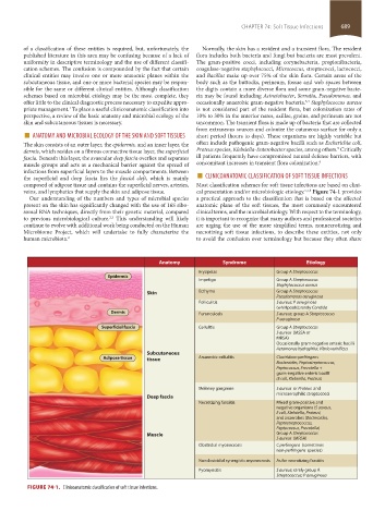
veins, and lymphatics that supply the skin and adipose tissue. cal presentation and/or microbiologic etiology. 5,7,8 Figure 74-1 provides
Our understanding of the numbers and types of microbial species a practical approach to the classification that is based on the affected
present on the skin has significantly changed with the use of 16S ribo- anatomic plane of the soft tissues, the most commonly encountered
somal RNA techniques, directly from their genetic material, compared clinical terms, and the microbial etiology. With respect to the terminology,
to previous microbiological culture. This understanding will likely it is important to recognize that many authors and professional societies
2,3
continue to evolve with additional work being conducted on the Human are urging the use of the more simplified terms, nonnecrotizing and
Microbiome Project, which will undertake to fully characterize the necrotizing soft tissue infections, to describe these entities, not only
human microbiota. 2 to avoid the confusion over terminology but because they often share
Anatomy Syndrome Etiology
Erysipelas Group A Streptococcus
Epidermis
Impetigo Group A Streptococcus
Staphylococcus aureus
Skin Ecthyma Group A Streptococcus
Pseudomonas aeruginosa
Folliculitis S aureus; P aeruginosa
(whirlpools); rarely Candida
Dermis Furunculosis S aureus; group A Streptococcus
P aeruginosa
Superficial fascia Cellulitis Group A Streptococcus
S aureus (MSSA or
MRSA)
Occasionally gram-negative enteric bacilli
Aeromonas hydrophila; Vibrio vulnificus
Subcutaneous
Adipose tissue tissue Anaerobic cellulitis Clostridium perfringens
Bacteroides, Peptostreptococcus,
Peptococcus, Prevotella +
gram-negative enteric bacilli
(E coli, Klebsiella, Proteus)
Meleney gangrene S aureus or Proteus and
microaerophilic streptococci
Deep fascia
Necrotizing fasciitis Mixed gram-positive and
negative organisms (S aureus,
E coli, Klebsiella, Proteus)
and anaerobes (Bacteroides,
Peptostreptococcus,
Peptococcus, Prevotella)
Muscle Group A Streptococcus
S aureus (MRSA)
Clostridial myonecrosis C perfringens (sometimes
non-perfringens species)
Nonclostridial synergistic myonecrosis As for necrotizing fasciitis
Pyomyositis S aureus; rarely group A
Streptococcus; P aeruginosa
FIGURE 74-1. Clinicoanatomic classification of soft tissue infections.
section05_c74-81.indd 689 1/23/2015 12:37:20 PM