
Page 113 - Clinical Anatomy
P. 113
ECA2 7/18/06 6:42 PM Page 98
98 The abdomen and pelvis
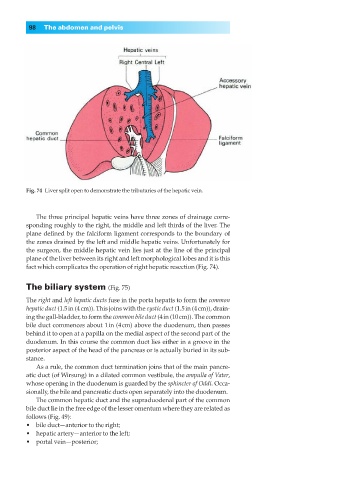
Fig. 74◊Liver split open to demonstrate the tributaries of the hepatic vein.
The three principal hepatic veins have three zones of drainage corre-
sponding roughly to the right, the middle and left thirds of the liver. The
plane defined by the falciform ligament corresponds to the boundary of
the zones drained by the left and middle hepatic veins. Unfortunately for
the surgeon, the middle hepatic vein lies just at the line of the principal
plane of the liver between its right and left morphological lobes and it is this
fact which complicates the operation of right hepatic resection (Fig. 74).
The biliary system (Fig. 75)
The right and left hepatic ducts fuse in the porta hepatis to form the common
hepatic duct (1.5in (4cm)). This joins with the cystic duct (1.5in (4cm)), drain-
ing the gall-bladder, to form the common bile duct (4in (10cm)). The common
bile duct commences about 1in (4cm) above the duodenum, then passes
behind it to open at a papilla on the medial aspect of the second part of the
duodenum. In this course the common duct lies either in a groove in the
posterior aspect of the head of the pancreas or is actually buried in its sub-
stance.
As a rule, the common duct termination joins that of the main pancre-
atic duct (of Wirsung) in a dilated common vestibule, the ampulla of Vater,
whose opening in the duodenum is guarded by the sphincter of Oddi. Occa-
sionally, the bile and pancreatic ducts open separately into the duodenum.
The common hepatic duct and the supraduodenal part of the common
bile duct lie in the free edge of the lesser omentum where they are related as
follows (Fig. 49):
•◊◊bile duct—anterior to the right;
•◊◊hepatic artery—anterior to the left;
•◊◊portal vein—posterior;