
Page 115 - Clinical Anatomy
P. 115
ECA2 7/18/06 6:42 PM Page 100
100 The abdomen and pelvis
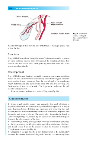
Fig. 76◊The arterial
supply of the gall-
bladder and Calot’s
triangle.
bladder through its bed directly into tributaries of the right portal vein
within the liver.
Structure
The gall-bladder wall and the sphincter of Oddi contain muscle, but there
are only scattered muscle fibres throughout the remaining biliary duct
system. The mucosa is lined throughout by columnar cells and bears
mucus-secreting glands.
Development
The gall-bladder and ducts are subject to numerous anatomical variations
which are best understood by considering their embryological develop-
ment. A diverticulum grows out from the ventral wall of the duodenum
which differentiates into the hepatic ducts and the liver (see Fig. 78).
Another diverticulum from the side of the hepatic duct bud forms the gall-
bladder and cystic duct.
Some variations are shown in a series of diagrams (Fig. 77).
Clinical features
1◊◊Errors in gall-bladder surgery are frequently the result of failure to
appreciate the variations in the anatomy of the biliary system; it is impor-
tant, therefore, before dividing any structures and removing the gall-
bladder, to have all the three biliary ducts clearly identified, together with
the cystic and hepatic arteries. The cystic artery is constantly found in
Calot’s triangle (Fig. 76), formed by the cystic duct, the common hepatic
duct and the inferior aspect of the liver.
2◊◊Haemorrhage during cholecystectomy may be controlled by compress-
ing the hepatic artery (which gives off the cystic branch) between the finger
and thumb where it lies in the anterior wall of the foramen of Winslow
(Pringle’s manoeuvre) (see Fig. 49).
3◊◊Gangrene of the gall-bladder is rare because even if the cystic artery
becomes thrombosed in acute cholecystitis there is a rich secondary blood