
Page 112 - Clinical Anatomy
P. 112
ECA2 7/18/06 6:42 PM Page 97
The gastrointestinal adnexae 97
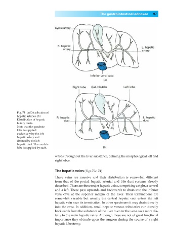
Fig. 73◊(a) Distribution of
hepatic arteries. (b)
Distribution of hepatic
biliary ducts.
Note that the quadrate
lobe is supplied
exclusively by the left
hepatic artery and
drained by the left
hepatic duct. The caudate
lobe is supplied by each.
wards throughout the liver substance, defining the morphological left and
right lobes.
The hepatic veins (Figs 72c, 74)
These veins are massive and their distribution is somewhat different
from that of the portal, hepatic arterial and bile duct systems already
described. There are three major hepatic veins, comprising a right, a central
and a left. These pass upwards and backwards to drain into the inferior
vena cava at the superior margin of the liver. Their terminations are
somewhat variable but usually the central hepatic vein enters the left
hepatic vein near its termination. In other specimens it may drain directly
into the cava. In addition, small hepatic venous tributaries run directly
backwards from the substance of the liver to enter the vena cava more dis-
tally to the main hepatic veins. Although these are not of great functional
importance they obtrude upon the surgeon during the course of a right
hepatic lobectomy.