
Page 116 - Clinical Anatomy
P. 116
ECA2 7/18/06 6:42 PM Page 101
The gastrointestinal adnexae 101
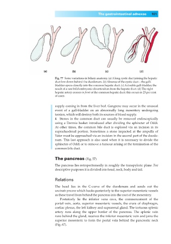
Fig. 77◊Some variations in biliary anatomy. (a) Along cystic duct joining the hepatic
duct low down behind the duodenum. (b) Absence of the cystic duct—the gall-
bladder opens directly into the common hepatic duct. (c) Adouble gall-bladder, the
result of a rare bifid embryonic diverticulum from the hepatic duct. (d) The right
hepatic artery crosses in front of the common hepatic duct; this occurs in 25 per cent
of cases.
supply coming in from the liver bed. Gangrene may occur in the unusual
event of a gall-bladder on an abnormally long mesentery undergoing
torsion, which will destroy both its sources of blood supply.
4◊◊Stones in the common duct can usually be removed endoscopically
using a Dormia basket introduced after dividing the sphincter of Oddi.
At other times, the common bile duct is explored via an incision in its
supraduodenal portion. Sometimes a stone impacted at the ampulla of
Vater must be approached via an incision in the second part of the duode-
num. This last approach is also used when it is necessary to divide the
sphincter of Oddi or to remove a tumour arising at the termination of the
common bile duct.
The pancreas (Fig. 57)
The pancreas lies retroperitoneally in roughly the transpyloric plane. For
descriptive purposes it is divided into head, neck, body and tail.
Relations
The head lies in the C-curve of the duodenum and sends out the
uncinate process which hooks posteriorly to the superior mesenteric vessels
as these travel from behind the pancreas into the root of the mesentery.
Posteriorly lie the inferior vena cava, the commencement of the
portal vein, aorta, superior mesenteric vessels, the crura of diaphragm,
coeliac plexus, the left kidney and suprarenal gland. The tortuous splenic
artery runs along the upper border of the pancreas. The splenic vein
runs behind the gland, receives the inferior mesenteric vein and joins the
superior mesenteric to form the portal vein behind the pancreatic neck
(Fig. 67).