
Page 170 - Clinical Anatomy
P. 170
ECA2 7/18/06 6:43 PM Page 155
The posterior abdominal wall 155
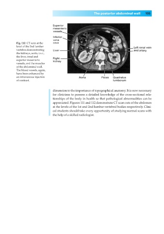
Fig. 112◊CT scan at the
level of the 2nd lumbar
vertebra demonstrating
the kidneys, aorta, i.v.c.,
the liver, renal and
superior mesenteric
vessels, and the muscles
of the abdominal wall.
The blood vessels, again,
have been enhanced by
an intravenous injection
of contrast.
dimension to the importance of topographical anatomy. It is now necessary
for clinicians to possess a detailed knowledge of the cross-sectional rela-
tionships of the body in health so that pathological abnormalities can be
appreciated. Figures 111 and 112 demonstrate CT scan cuts of the abdomen
at the levels of the 1st and 2nd lumbar vertebral bodies respectively. Clini-
cal students should take every opportunity of studying normal scans with
the help of a skilled radiologist.